Clinical Trial
. 2012;7(2):e30037.
doi: 10.1371/journal.pone.0030037. Epub 2012 Feb 17.

Amplitude reduction and phase shifts of melatonin, cortisol and other circadian rhythms after a gradual advance of sleep and light exposure in humans

Affiliations
Clinical Trial

Amplitude reduction and phase shifts of melatonin, cortisol and other circadian rhythms after a gradual advance of sleep and light exposure in humans

Derk-Jan Dijk et al. PLoS One. 2012.

Abstract

Background: The phase and amplitude of rhythms in physiology and behavior are generated by circadian oscillators and entrained to the 24-h day by exposure to the light-dark cycle and feedback from the sleep-wake cycle. The extent to which the phase and amplitude of multiple rhythms are similarly affected during altered timing of light exposure and the sleep-wake cycle has not been fully characterized.

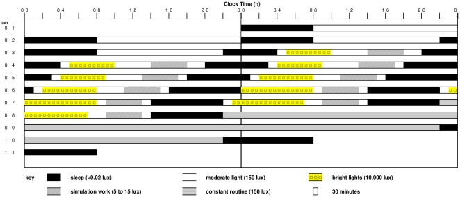
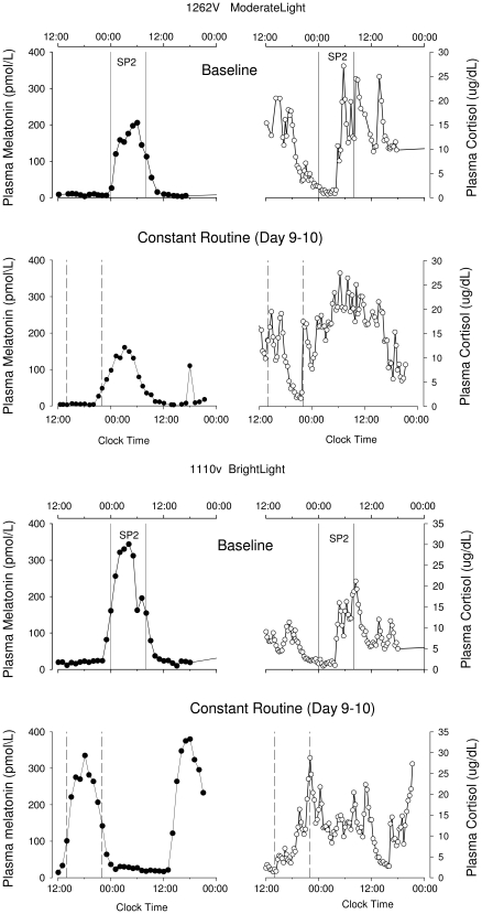
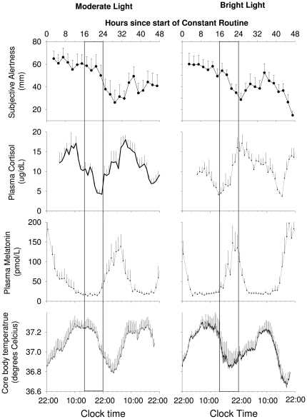
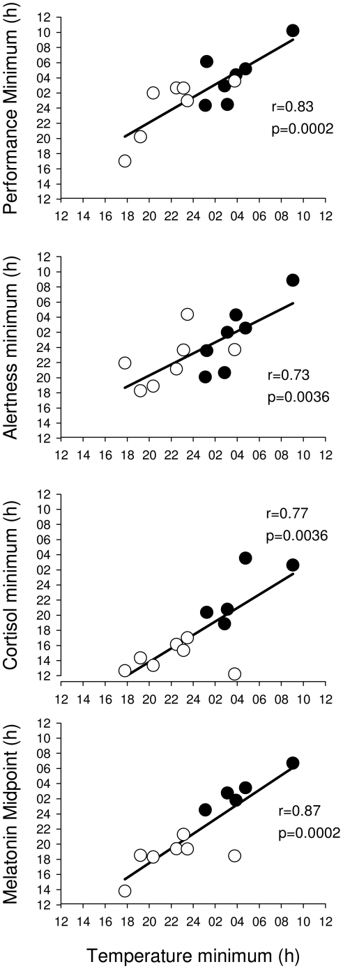
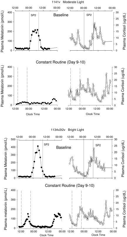
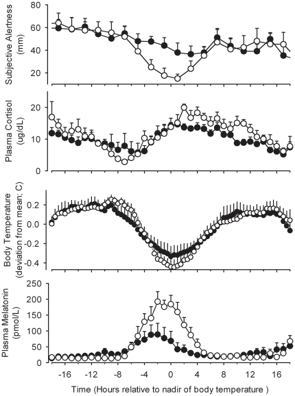
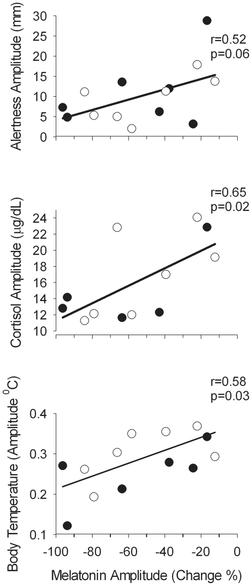
Methodology/principal findings: We assessed the phase and amplitude of the rhythms of melatonin, core body temperature, cortisol, alertness, performance and sleep after a perturbation of entrainment by a gradual advance of the sleep-wake schedule (10 h in 5 days) and associated light-dark cycle in 14 healthy men. The light-dark cycle consisted either of moderate intensity 'room' light (∼90-150 lux) or moderate light supplemented with bright light (∼10,000 lux) for 5 to 8 hours following sleep. After the advance of the sleep-wake schedule in moderate light, no significant advance of the melatonin rhythm was observed whereas, after bright light supplementation the phase advance was 8.1 h (SEM 0.7 h). Individual differences in phase shifts correlated across variables. The amplitude of the melatonin rhythm assessed under constant conditions was reduced after moderate light by 54% (17-94%) and after bright light by 52% (range 12-84%), as compared to the amplitude at baseline in the presence of a sleep-wake cycle. Individual differences in amplitude reduction of the melatonin rhythm correlated with the amplitude of body temperature, cortisol and alertness.

Conclusions/significance: Alterations in the timing of the sleep-wake cycle and associated bright or moderate light exposure can lead to changes in phase and reduction of circadian amplitude which are consistent across multiple variables but differ between individuals. These data have implications for our understanding of circadian organization and the negative health outcomes associated with shift-work, jet-lag and exposure to artificial light.


Conflict of interest statement

Competing Interests: Dr. Dijk has received research funding from GSK, Eli Lilly, Merck, Ono Pharma, Philips, Airforce Office of Scientific Research, Biotechnology and the Biological Sciences Research Council and provided consulting services to Johnson and Johnson, Sanofi-aventis, Metro Naps, Pepsi Co, Actelion, Cephalon, GSK, Eli Lilly, Philips, Merck, Ono Pharma, UCB, and CHDI. Dr. Czeisler has received consulting fees/served as a paid member of scientific advisory boards for: Actelion, Ltd.; Avera Pharmaceuticals, Inc.; Bombardier Inc.; Boston Celtics; Cephalon, Inc.; Columbia River Bar Pilots; Delta Airlines; Eli Lilly and Co.; Federal Motor Carrier Safety Administration (FMCSA), U.S. Department of Transportation; Fedex Kinko's; Fusion Medical Education, LLC; Garda Síochána Inspectorate; Global Ground Support; Hypnion, Inc. (acquired by Eli Lilly and Co. in April 2007); Johnson & Johnson; Koninklijke Philips Electronics, N.V.; Minnesota Timberwolves; Morgan Stanley; Norfolk Southern; Portland Trail Blazers; Respironics, Inc.; Sanofi-Aventis Groupe; Sepracor, Inc.; Sleep Multimedia, Inc.; Sleep Research Society; Somnus Therapeutics, Inc.; Takeda Pharmaceuticals; Vanda Pharmaceuticals, Inc.; Vital Issues in Medicine; Warburg-Pincus;Zeo Inc. Dr. Czeisler owns an equity interest in Lifetrac, Inc.; Somnus Therapeutics, Inc.; Vanda Pharmaceuticals, Inc., and Zeo Inc. Dr. Czeisler received royalties from the Massachusetts Medical Society/New England Journal of Medicine; McGraw Hill, the New York Times Penguin Press and Philips Respironics. Dr. Czeisler has clinical trial research contracts from Cephalon, Inc., Merck & Co., Inc., and Pfizer, Inc.; an investigator-initiated research grant from Cephalon, Inc.; and his research laboratory at the Brigham and Women's Hospital has received unrestricted research and education funds for research expenses from Cephalon, Inc., Koninklijke Philips Electronics, N.V., ResMed, and the Brigham and Women's Hospital. Dr. Czeisler is the incumbent of an endowed professorship provided to Harvard University by Cephalon, Inc. and holds a number of process patents in the field of sleep/circadian rhythms,details of which are available on request. Since 1985, Dr. Czeisler has also served as an expert witness on various legal cases related to sleep and/or circadian rhythms. Dr. Duffy has received investigator-initiated research grants from Philips-Respironics. Dr. Boivin has received research support from Transport Canada and the Litebook Company; has participated in speaking and advisory engagements for Servier and Cephalon and is Founder/CEO of Alpha Logik Consultants, Inc. Dr. Shanahan, declares that her spouse is Dr. Czeisler. None of the commercial research grants listed in the statement relate to the current study. Our listed conflicts do not alter our adherence to all the PLoS ONE policies on sharing data and materials, as detailed online in the guide for authors.

Figures

Figure 1
Figure 1. Raster plot of the experimental protocol.
During the two baseline days sleep episodes were scheduled from 00:00 to 08:00 Thereafter, the sleep-wake schedule was advanced gradually resulting in a 10 hour advance of the sleep-wake schedule. Sleep episodes on day seven and eight were scheduled from 14:00 to 22.00. During scheduled sleep episodes, participants were exposed to darkness (<0.02 lux). During waking hours the participants were exposed to ∼90–150 lux moderate light. During the bright light treatment episodes, the moderate light participants remained in moderate light, whereas the bright light treatment participants received ∼10,000 lux of light. Both moderate light and bright light treated participants participated in dim light work simulations.
Figure 2
Figure 2. Plasma melatonin and cortisol rhythms.
Plasma melatonin and cortisol rhythms before (Baseline – SP2 – upper panels) and after (Constant Routine – lower panels) the gradual shift of the sleep-wake schedule, in a participant treated with moderate light (1262V) and a participant treated with bright light (1110V). The vertical solid lines indicate the eight-hour scheduled sleep/darkness episode (SP2) on the baseline day (D2), the dotted vertical lines indicate the projected eight hour sleep episode (based on the sleep period of the preceding night) during the constant routine.
Figure 3
Figure 3. Average waveforms of circadian variables after moderate and bright light treatment.
Time course of core body temperature, plasma melatonin, cortisol and subjective alertness during the constant routine in moderate light and bright light treated participants. Subjective alertness data were averaged per 2-h bins. All date are referenced to the projected wake time (22:00 clock-time). Box indicates the timing of the sleep episode on the previous day. Data represent means+/− SEMs.
Figure 4
Figure 4. Association between the timing of circadian variables.
Association between the timing of melatonin midpoint, cortisol minimum, circadian minimum of alertness and performance and the timing of the temperature minimum during the constant routine in participants exposed to moderate light (filled symbols) or bright light (open symbols).
Figure 5
Figure 5. Examples of amplitude reduction.
The plasma melatonin (filled symbols) and cortisol rhythm (open symbols) during the baseline day and SP2 and the constant routine (Day 9–10) in a bright light (1134) and moderate light (1141) treated subject. In both participants a reduction of the plasma melatonin amplitude as well as changes in the cortisol rhythm can be observed.
Figure 6
Figure 6. Average waveforms of circadian variables and amplitude reduction.
Average waveform of subjective alertness, plasma cortisol, core body temperature and plasma melatonin during a constant routine in participants with a reduction in melatonin amplitude >50% (closed symbols; n = 7) compared to those in whom the reduction was <50% (open symbols; n = 7). All data are aligned with the timing of the fitted minimum of the core body temperature rhythm. Error bars indicate 1 SEM.
Figure 7
Figure 7. Association between the amplitude of circadian variables.
Association between the amplitude of body temperature, cortisol, and alertness during the constant routine and the reduction in the amplitude of melatonin in participants exposed to moderate light (filled symbols) or bright light (open symbols).

Similar articles

Cited by

References

    1. Czeisler CA, Dijk D-J. Human circadian physiology and sleep-wake regulation. In: Takahashi JS, Turek FW, Moore RY, editors. Handbook of Behavioral Neurobiology: Circadian Clocks. New York: Kluwer Academic/Plenum Publishing Co; 2001. pp. 531–561.
    1. Czeisler CA, Gooley JJ. Sleep and circadian rhythms in humans. Cold Spring Harb Symp Quant Biol. 2007;72:579–597. - PubMed
    1. Takahashi JS, Hong HK, Ko CH, McDearmon EL. The genetics of mammalian circadian order and disorder: implications for physiology and disease. Nat Rev Genet. 2008;9:764–775. - PMC - PubMed
    1. Welsh DK, Logothetis DE, Meister M, Reppert SM. Individual neurons dissociated from rat suprachiasmatic nucleus express independently phased circadian firing rhythms. Neuron. 1995;14:697–706. - PubMed
    1. Guler AD, Ecker JL, Lall GS, Haq S, Altimus CM, et al. Melanopsin cells are the principal conduits for rod-cone input to non-image-forming vision. Nature. 2008;453:102–105. - PMC - PubMed

Publication types