. 2012;7(2):e30210.
doi: 10.1371/journal.pone.0030210. Epub 2012 Feb 17.

Novel transgenic mice for inducible gene overexpression in pancreatic cells define glucocorticoid receptor-mediated regulations of beta cells

Affiliations

Novel transgenic mice for inducible gene overexpression in pancreatic cells define glucocorticoid receptor-mediated regulations of beta cells

Bertrand Blondeau et al. PLoS One. 2012.

Abstract

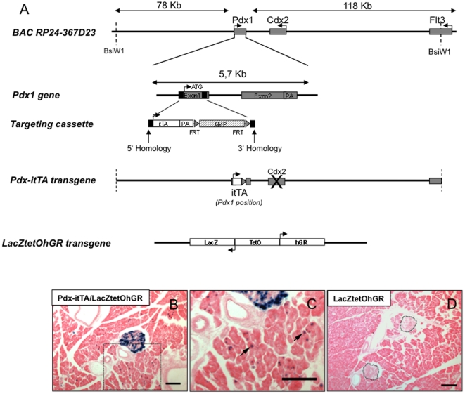
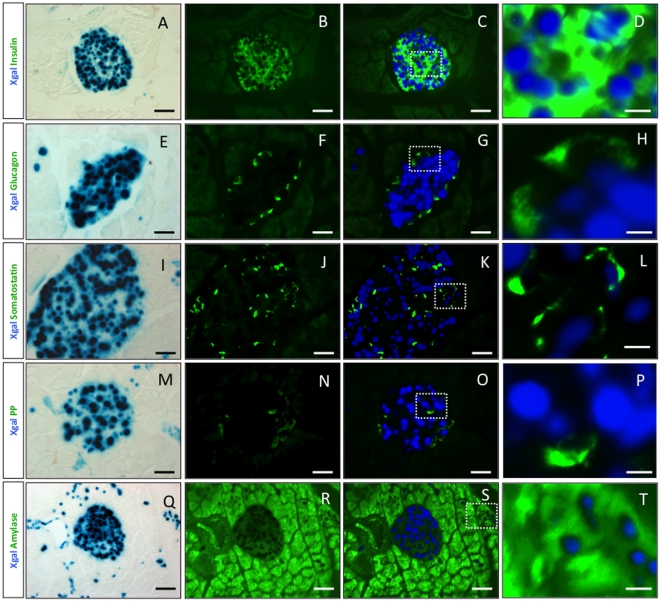
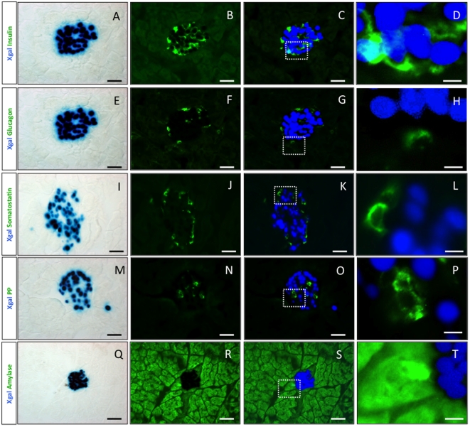
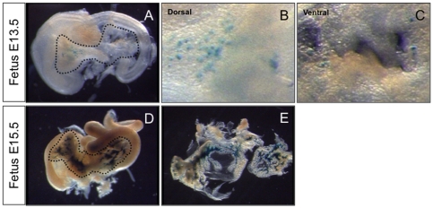
Conditional gene deletion in specific cell populations has helped the understanding of pancreas development. Using this approach, we have shown that deleting the glucocorticoid receptor (GR) gene in pancreatic precursor cells leads to a doubled beta-cell mass. Here, we provide genetic tools that permit a temporally and spatially controlled expression of target genes in pancreatic cells using the Tetracycline inducible system. To efficiently target the Tetracycline transactivator (tTA) in specific cell populations, we generated Bacterial Artificial Chromosomes (BAC) transgenic mice expressing the improved Tetracycline transactivator (itTA) either in pancreatic progenitor cells expressing the transcription factor Pdx1 (BAC-Pdx1-itTA), or in beta cells expressing the insulin1 gene (BAC-Ins1-itTA). In the two transgenic models, itTA-mediated activation of reporter genes was efficient and subject to regulation by Doxycycline (Dox). The analysis of a tetracycline-regulated LacZ reporter gene shows that in BAC-Pdx1-itTA mice, itTA is expressed from embryonic (E) day 11.5 in all pancreatic precursor cells. In the adult pancreas, itTA is active in mature beta, delta cells and in few acinar cells. In BAC-Ins1-itTA mice tTA is active from E13.5 and is restricted to beta cells in fetal and adult pancreas. In both lines, tTA activity was suppressed by Dox treatment and re-induced after Dox removal. Using these transgenic lines, we overexpressed the GR in selective pancreatic cell populations and found that overexpression in precursor cells altered adult beta-cell fraction but not glucose tolerance. In contrast, GR overexpression in mature beta cells did not alter beta-cell fraction but impaired glucose tolerance with insufficient insulin secretion. In conclusion, these new itTA mouse models will allow fine-tuning of gene expression to investigate gene function in pancreatic biology and help us understand how glucocorticoid signaling affects on the long-term distinct aspects of beta-cell biology.


Conflict of interest statement

Competing Interests: The authors have declared that no competing interests exist.

Figures

Figure 1
Figure 1. Generation and characterization of mice expressing the itTA under the control of Pdx1 regulatory elements.
(A) Bacterial Artificial Chromosomes (BAC) containing 202 kb of the genomic region of Pdx1 was obtained from a BAC library developed by the CHORI, Oakland, USA. The coding region of the gene was removed by homologous recombination in bacteria and replaced by the itTA cDNA. The construct was then injected in the pronuclei of fertilized eggs. itTA = improved tetracycline transactivator; AMP = ampicillin resistance gene; PA = polyadenylation site; FRT = Flippase recognition target. Below, a scheme representing the LacZtetOhGR construct (B) Lac Z expression revealed by Xgal staining (blue) in islets and in exocrine tissue in Pdx-itTA/LacZtetOhGR mice. (C) Magnified view of the boxed area of B with arrows pointing at blue cells in the exocrine tissue. (D) Absence of blue staining in control mice carrying only the LacZtetOhGR transgene. Two islets are outlined. Scale bar = 50 µm.
Figure 2
Figure 2. The Pdx1-itTA activates lacZ expression in beta, delta and in acinar cells.
(A) (E) (I) (M) and (Q) LacZ expression revealed by Xgal staining (blue) on adult pancreatic sections from a Pdx1-itTA/LacZtetOhGR mouse. (B) Immunofluorescence for insulin (green), (C) merge of A and B and (D) magnified view of inset in C. (F) Immunofluorescence for glucagon (green), (G) merge of E and F and (H) magnified view of inset in G. (J) Immunofluorescence for somatostatin (green), (K) merge of I and J and (L) magnified view of inset in K. (N) Immunofluorescence for PP (green), (O) merge of M and N and (P) magnified view of inset in O. (R) Immunofluorescence for amylase (green), (S) merge of Q and R and (T) magnified view of inset in S. Scale bar = 50 µm except for D, H, L, P and T where scale bar = 10 µm.
Figure 3
Figure 3. The Pdx1-itTA is active early during fetal development in pancreatic precursors.
(A) LacZ expression revealed by Xgal staining in pancreatic buds at E11.5 and in the entire pancreas at E13.5 (B) and E15.5 (C) from Pdx-itTA/LacZtetOhGR fetuses. d = dorsal; v = ventral; st = stomach.
Figure 4
Figure 4. Generation and characterization of mice expressing the itTA under the control of Insulin1 regulatory elements.
(A) Bacterial Artificial Chromosomes (BAC) containing around 200 kb of the genomic region of Insulin1 was obtained from BAC libraries developed by the CHORI, Oakland USA. The coding region of the gene was removed by homologous recombination in bacteria and replaced by the itTA cDNA. The construct was then injected in the pronuclei of fertilized eggs. itTA = humanized tetracycline transactivator; AMP = ampicillin resistance gene; PA = polyadenylation site; FRT = Flippase recognition target. (B) Lac Z expression revealed by Xgal staining (blue) in islets in Ins1-itTA/LacZtetOhGR mice. (C) Absence of blue staining in control mice carrying only the LacZtetOhGR transgene. Two islets are outlined. Scale bar = 50 µm.
Figure 5
Figure 5. The Ins1-itTA transgene activates lacZ expression only in beta cells.
(A), (E), (I), (M) and (Q) LacZ expression revealed by Xgal staining (blue) on adult pancreatic sections from a Ins1-itTA/LacZtetOhGR mouse. (B) Immunofluorescence for insulin (green), (C) merge of A and B and (D) magnified view of inset in C. (F) Immunofluorescence for glucagon (green), (G) merge of E and F and (H) magnified view of inset in G. (J) Immunofluorescence for somatostatin (green), (K) merge of I and J and (L) magnified view of inset in K. (N) Immunofluorescence for PP (green), (O) merge of M and N and P magnified view of inset in O. (R) Immunofluorescence for amylase (green), (S) merge of Q and R and (T) magnified view of inset in S. Scale bar = 50 µm except for D, H, L, P and T where scale bar = 10 µm.
Figure 6
Figure 6. The Ins1-itTA is active in cells expressing insulin during fetal life.
(A) LacZ expression in pancreatic buds at E13.5 in the dorsal (B) and ventral (C) part of the pancreas from Ins1-itTA/LacZtetOhGR fetuses. (D) LacZ expression is found at E15.5 in scattered cells. (E) A view of the dissected pancreas at E15.5.
Figure 7
Figure 7. The expression of LacZ can be extinguished or induced in adult Pdx1-itTA/LacZtetOhGR mice.
(A) Xgal staining on a pancreatic section of an adult Pdx1-itTA/LacZtetOhGR mouse without Dox treatment. (B) Absence of Xgal staining after 8 weeks of Dox administration (1 mg/ml) in the drinking water of an adult Pdx1-itTA/LacZtetOhGR animal. An islet is outlined. (C) Absence of Xgal staining in a 28 days-old Pdx1-itTA/LacZtetOhGR mouse treated with Dox (0.1 mg/ml) from E0. An islet is outlined. (D) Eight weeks after Dox removal, Xgal staining is observed on a pancreatic section from a Pdx1-itTA/LacZtetOhGR mouse. Note that in contrast with A, Xgal staining is observed in islet cells and not in acinar cells. Scale bar = 50 µm.
Figure 8
Figure 8. Consequences of GR overexpression on beta-cell mass and glucose homeostasis in Pdx1-itTA/LacZtetOhGR or Ins1-itTA/LacZtetOhGR mice.
(A) Immunohistochemistry for the glucocorticoid receptor (GR, brown nuclear staining) on pancreatic sections from a control LacZtetOhGR, (B) a Pdx1-itTA/LacZtetOhGR mouse and (C) a Ins1-itTA/LacZtetOhGR mouse. (D) Beta-cell fraction, (E) beta-cell mass, (F) blood glucose during an intra-peritoneal glucose tolerance test and (G) corresponding Area Under the Curve (AUC) in adult Pdx1-itTA/LacZtetOhGR mice. (H) Beta-cell fraction, (I) beta-cell mass, (J) blood glucose during an intra-peritoneal glucose tolerance test and (K) corresponding Area Under the Curve (AUC) in adult Ins1-itTA/LacZtetOhGR mice. Results are expressed as means ± SD for n = 3–5 animals per group. * p<0.05 when comparing double transgenic mice (Pdx1-itTA/LacZtetOhGR or Ins1-itTA/LacZtetOhGR) to control (LacZtetOhGR) mice using a Mann-Whitney non parametric test. Scale bar = 100 µm.

Similar articles

Cited by

References

    1. Slack JM. Developmental biology of the pancreas. Development. 1995;121:1569–1580. - PubMed
    1. Murtaugh LC, Melton DA. Genes, signals, and lineages in pancreas development. Annu Rev Cell Dev Biol. 2003;19:71–89. - PubMed
    1. Gesina E, Tronche F, Herrera P, Duchene B, Tales W, et al. Dissecting the role of glucocorticoids on pancreas development. Diabetes. 2004;53:2322–2329. - PubMed
    1. Blondeau B, Lesage J, Czernichow P, Dupouy JP, Breant B. Glucocorticoids impair fetal beta-cell development in rats. Am J Physiol Endocrinol Metab. 2001;281:E592–E599. - PubMed
    1. Valtat B, Dupuis C, Zenaty D, Singh-Estivalet A, Tronche F, et al. Genetic evidence of the programming of beta cell mass and function by glucocorticoids in mice. Diabetologia. 2011;54:350–359. - PubMed

Publication types

LinkOut - more resources