. 2012;7(2):e30562.
doi: 10.1371/journal.pone.0030562. Epub 2012 Feb 20.

CCN2/connective tissue growth factor is essential for pericyte adhesion and endothelial basement membrane formation during angiogenesis

Affiliations

CCN2/connective tissue growth factor is essential for pericyte adhesion and endothelial basement membrane formation during angiogenesis

Faith Hall-Glenn et al. PLoS One. 2012.

Abstract

CCN2/Connective Tissue Growth Factor (CTGF) is a matricellular protein that regulates cell adhesion, migration, and survival. CCN2 is best known for its ability to promote fibrosis by mediating the ability of transforming growth factor β (TGFβ) to induce excess extracellular matrix production. In addition to its role in pathological processes, CCN2 is required for chondrogenesis. CCN2 is also highly expressed during development in endothelial cells, suggesting a role in angiogenesis. The potential role of CCN2 in angiogenesis is unclear, however, as both pro- and anti-angiogenic effects have been reported. Here, through analysis of Ccn2-deficient mice, we show that CCN2 is required for stable association and retention of pericytes by endothelial cells. PDGF signaling and the establishment of the endothelial basement membrane are required for pericytes recruitment and retention. CCN2 induced PDGF-B expression in endothelial cells, and potentiated PDGF-B-mediated Akt signaling in mural (vascular smooth muscle/pericyte) cells. In addition, CCN2 induced the production of endothelial basement membrane components in vitro, and was required for their expression in vivo. Overall, these results highlight CCN2 as an essential mediator of vascular remodeling by regulating endothelial-pericyte interactions. Although most studies of CCN2 function have focused on effects of CCN2 overexpression on the interstitial extracellular matrix, the results presented here show that CCN2 is required for the normal production of vascular basement membranes.


Conflict of interest statement

Competing Interests: The authors have declared that no competing interests exist.

Figures

Figure 1
Figure 1. Expression of Ccn2 in developing vasculature.
(A) β-galactosidase activity in Ccn2-lacZ transgenic mice reveals Ccn2 promoter activity throughout the vasculature in E16.5 embryos. (B) Ccn2-lacZ expression in dermal microvessels at E13.5. (C–E) EGFP fluorescence in CCN2-EGFP BAC transgenic mice demonstrates CCN2 expression in the endothelium of arterial elements (C and E), venous elements (C), and developing capillary networks (D). Arrowheads in (C) and (E) demarcate arterial element. Arrow in (C) identifies endothelial cells of a venous element. Arrowhead in (E) highlights EGFP expression in mural cells in the arterial element. Arrow in (E) highlights expression in endothelial cells in the arterial element. (F) Immunofluorescence and (G,H) immunohistochemical staining with an αCCN2 antibody on paraffin sections through dermis, demonstrating CCN2 expression in endothelial cells. Arrows in (F) highlight endothelial cells in E18.5 microvasculature. Specificity of the αCCN2 antibody is demonstrated by the absence of reactivity in the Ccn2−/− section (H). Arrows in (G) and (H) demarcate abluminal surface of the endothelium. Asterisks in (G) and (H) identify blood cells within the vessels. αCCN2 staining in (G) shows punctate intracellular expression, presumably with the Golgi, in addition to the surface expression marked by the arrow.
Figure 2
Figure 2. Vascular abnormalities in Ccn2 mutant embryos.
(A) E18.5 WT and (B) Ccn2−/− littermate, showing vessel dilation throughout the mutant embryo. (C, D) H&E-stained paraffin sections through the lumbar dorsal dermis of (C) E18.5 WT and (D) Ccn2−/− littermate. Arrowheads point to vessels. Bars highlight the enlarged distance between the hypodermal and epidermal layers in the mutant, indicative of local edema. (E,F) Hematoxylin and eosin-stained sections through E16.5 WT (E) and Ccn2−/− (F) descending aorta at thoracic level. Smooth muscle cells in the tunica media are spindle-shaped and arranged in layers in the WT embryo, but are more cuboidal and disorganized in the Ccn2−/− littermate. (G,H) Higher magnification images through aorta at lumbar level in E16.5 (G) WT and (H) Ccn2−/− littermate showing spindle-shaped smooth muscle cells (arrowheads) in WT that have a cuboidal shape in the mutant. (I,J) Confocal images of PECAM-stained dorsal dermal vasculature in (I) WT and (J) Ccn2−/− littermates. Arrows demarcate arterial elements; arrowheads demarcate venous elements; asterisks identify capillary beds. (K,L) Higher magnification confocal images of (K) WT and (L) Ccn2−/− dorsal dermal capillary beds, showing increased capillary density in the mutant. (M,N) High magnification confocal image of (M) WT and (N) Ccn2−/− dorsal dermal capillaries, showing numerous abluminal protrusions (arrows in (N)) on the mutant capillary. (O,P) Electron micrographs of newborn (P0) (O) WT and (P) Ccn2−/− dermal capillaries, showing abluminal and luminal (arrows in (P)) protrusions.
Figure 3
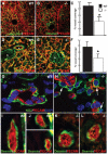
Figure 3. Defective endothelial-pericyte interactions in Ccn2 mutants.
(A, B) Co-immunofluorescence staining for desmin and PECAM in E18.5 dermis from (A) WT and (B) Ccn2−/− mice analyzed by confocal microscopy. (C) Quantification of vessel coverage by pericytes in E18.5 dermis; asterisk, p<0.05. (D, E) Co-immunofluorescence staining for NG2 and PECAM in E16.5 lung from (D) WT and (E) Ccn2−/− mice analyzed by confocal microscopy. (F) Quantification of vessel coverage by pericytes in E16.5 lung; asterisk, p<0.05. (G,H) Confocal analysis of NG2 and PECAM immunostaining in (G) WT and (H) Ccn2−/− E16.5 dermis. Pericytes are elongated around the microvessel in (G), whereas in mutants (H), pericytes (arrows) are associated with the endothelium, but are rounder, and fewer of them have elongated along the endothelial surface. (I–L) Confocal sections through E16.5 dermis analyzed for desmin (green) and PECAM (red) immunofluorescence. (I,J) WT desmin positive pericytes appear elongated and cover most of the surface of the microvessels. (K.L) Ccn2−/− desmin-positive pericytes have a rounder appearance and show less extensive coverage of the surface of the endothelium.
Figure 4
Figure 4. CCN2 potentiates PDGF-B signaling.
(A) rCCN2 induces PDGF-B expression in HUVEC cells. Right panel, representative Western blot. Left panel, Quantification of relative expression levels of PDGF-B in cells treated with or without rCCN2 from three separate experiments. *, p<0.02. (B) Adenovirally expressed CCN2 induces PDGF-B expression in HUVECs compared to transfection with an empty adenoviral control. The extent of PDGF-B induction correlated with levels of CCN2 expression. As reported previously, a higher molecular weight isoform of CCN2, presumably a result of post-translational modification , is detected 4 and 8 hours post-infection. Relative level of PDGF-B expression was assessed using ImageJ software. The experiment was repeated three times, with similar results each time. The induction of PDGF-B in the presence of CCN2 was statistically significant for each time point; p<0.05. A representative Western blot is shown. (C) Effects of rPDGF-B, and/or pcDNA3-CCN2-HA expression on activation of PDGF pathways in MOVAS cells. PDGF-B stimulated activation of Stat3, ERK, and Akt, whereas CCN2-HA on its own had no effect. However, combined treatment with PDGF-B and CCN2-HA led to prolonged Akt activation (arrows). Relative levels of pAKT expression were assessed using ImageJ software. All experiments were performed in triplicate and repeated three times, with similar results each time. The increase in pAKT levels in the presence of CCN2 was statistically significant at each time point; p<0.05. A representative Western blot is shown.
Figure 5
Figure 5. Endothelial basement membrane defects in Ccn2 mutants.
Electron microcopic images of endothelial basement membranes in dermal capillaries of E16.5 (A) WT and (B) Ccn2−/− littermates. Arrows demarcate the plasma membrane (bottom arrow) and top of the interstitial matrix (top arrow). (C,D) Confocal images of dermis of E16.5 WT (C) and Ccn2−/− (D) mice analyzed by immunofluorescence for fibronectin (FN) and PECAM. Arrows identify an arteriole. The arteriole in (C) is surrounded by several layers of FN. The arteriole in (D) is incompletely invested with FN. (E,F), Lower magnification confocal images through (E) WT and (F) Ccn2−/− E16.5 dermis, illustrating less fibronectin throughout the dermis in mutants. (G,H) Confocal images of dermis of E16.5 (G) WT and (H) Ccn2−/− mice analyzed by immunofluorescence for ColIV (Col4α2) and PECAM. Arrows identify an arteriole. ColIV coverage of the mutant vasculature is incomplete. (I,J) Confocal images of lungs of E16.5 (I) WT and (J) Ccn2−/− mice analyzed by immunofluorescence for ColIV and PECAM. Most of the vascular elements in the WT lung are surrounded by ColIV. Coverage is incomplete in the Ccn2 mutant lung. Arrows in (J) identify vessels lacking coverage by ColIV. (K) CCN2 induces expression of FN and ColIV in HUVECS. HUVECs were infected with Ad-CCN2-GFP or Ad-control. Lysates were collected at the indicated time points post-infection. Levels of FN are elevated 8 hours after infection, concomitant with accumulation of CCN2. There appeared to be an increase in FN levels at 12 hours in the presence of CCN2 in the blot shown, but this was not seen in every experiment and the result did not reach statistical significance at this time point. Similarly, there was a trend towards increased expression of Col IV at 12 hr, but this increase did not reach statistical significance (p = 0.065). The experiment was repeated three times. A representative blot is shown. Quantification of levels of FN and Col IV are shown in Figure S6.

Similar articles

Cited by

References

    1. Brigstock DR. The CCN family: a new stimulus package. J Endocrinol. 2003;178:169–175. - PubMed
    1. Bork P. The modular architecture of a new family of growth regulators related to connective tissue growth factor. FEBS Lett. 1993;327:125–130. - PubMed
    1. Vorwerk P, Hohmann B, Oh Y, Rosenfeld RG, Shymko RM. Binding properties of insulin-like growth factor binding protein-3 (IGFBP-3), IGFBP-3 N- and C-terminal fragments, and structurally related proteins mac25 and connective tissue growth factor measured using a biosensor. Endocrinology. 2002;143:1677–1685. - PubMed
    1. Abreu JG, Ketpura NI, Reversade B, De Robertis EM. Connective-tissue growth factor (CTGF) modulates cell signalling by BMP and TGF-beta. Nat Cell Biol. 2002;4:599–604. - PMC - PubMed
    1. Hashimoto G, Inoki I, Fujii Y, Aoki T, Ikeda E, et al. Matrix metalloproteinases cleave connective tissue growth factor and reactivate angiogenic activity of vascular endothelial growth factor 165. J Biol Chem. 2002;277:36288–36295. - PubMed

Publication types

MeSH terms

Substances