. 2012;7(2):e30563.
doi: 10.1371/journal.pone.0030563. Epub 2012 Feb 20.

Origins of the tumor microenvironment: quantitative assessment of adipose-derived and bone marrow-derived stroma

Affiliations

Origins of the tumor microenvironment: quantitative assessment of adipose-derived and bone marrow-derived stroma

Shannon Kidd et al. PLoS One. 2012.

Abstract

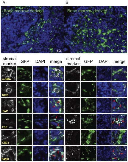
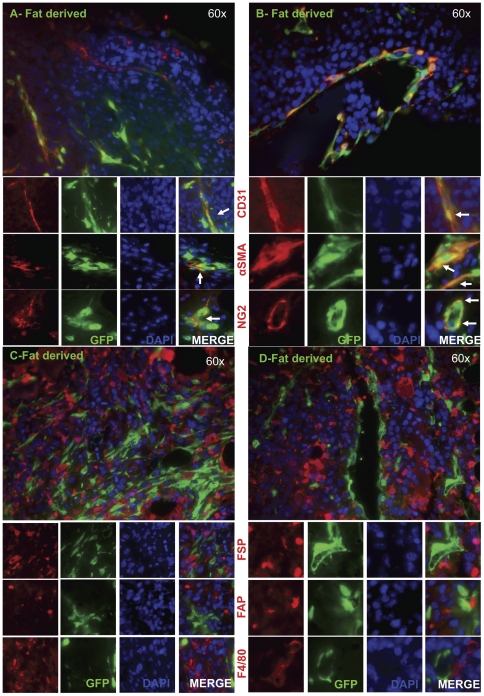
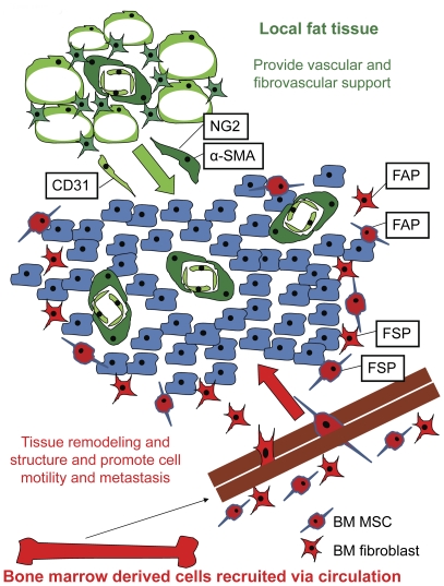
To meet the requirements for rapid tumor growth, a complex array of non-neoplastic cells are recruited to the tumor microenvironment. These cells facilitate tumor development by providing matrices, cytokines, growth factors, as well as vascular networks for nutrient and waste exchange, however their precise origins remain unclear. Through multicolored tissue transplant procedures; we have quantitatively determined the contribution of bone marrow-derived and adipose-derived cells to stromal populations within syngeneic ovarian and breast murine tumors. Our results indicate that subpopulations of tumor-associated fibroblasts (TAFs) are recruited from two distinct sources. The majority of fibroblast specific protein (FSP) positive and fibroblast activation protein (FAP) positive TAFs originate from mesenchymal stem/stromal cells (MSC) located in bone marrow sources, whereas most vascular and fibrovascular stroma (pericytes, α-SMA(+) myofibroblasts, and endothelial cells) originates from neighboring adipose tissue. These results highlight the capacity for tumors to utilize multiple sources of structural cells in a systematic and discriminative manner.


Conflict of interest statement

Competing Interests: The authors have declared that no competing interests exist.

Figures

Figure 1
Figure 1. Bone marrow tissue contributions to tumor stromal elements.
Lethally irradiated RFP+ mice were reconstituted with GFP+ bone marrow (n = 3). After engraftment, ID8 cells were injected subcutaneously. 5 weeks later, tumors were harvested, and sections were analyzed for α-SMA, NG2, FAP, FSP, CD31, and F4/80 co-staining with GFP+ bone marrow derived cells or RFP+ non bone marrow derived cells. (A&B) Red arrows represent co-staining of GFP+ bone marrow derived cells with FAP, FSP or F4/80. Representative images are shown for 2 separate animals.
Figure 2
Figure 2. Non-bone marrow tissue contributions to tumor stromal elements.
Lethally irradiated RFP+ mice were reconstituted with GFP+ bone marrow (n = 3). After engraftment, ID8 cells were injected subcutaneously. 5 weeks later, tumors were harvested, and sections were analyzed for α-SMA, NG2, FAP, FSP, CD31, and F4/80 co-staining with GFP+ bone marrow derived cells or RFP+ non bone marrow derived cells. (A&B) Yellow arrows represent co-staining of RFP+ non-bone marrow derived cells with α-SMA, NG2 or CD31. Representative images are shown for 2 separate animals.
Figure 3
Figure 3. BM MSC contribution to the tumor microenvironment.
Prospectively isolated and in vitro isolated MSC (RFP+) were combined with whole marrow and transplanted into lethally irradiated mice (n = 3 for each of the 2 groups). After engraftment, ID8 cells were subcutaneously injected. After 5 weeks of tumor groups, sections from harvested tumors were analyzed for RFP+ cells. (A) Representative images are shown for RFP co-staining with FAP and FSP as indicated by the white arrows in the merged images. There is no co-staining between RFP+ cells and α-SMA. (B) Scores were assessed for the relative percentage of RFP+ cells co-staining with each stromal marker.
Figure 4
Figure 4. Engrafted GFP+ adipose tissue is locally recruited into the tumor microenvironment.
GFP+ adipose tissue was subcutaneously implanted into wild type mice (n = 3). After engraftment, E0771 cells were subcutaneously injected. Two weeks later, sections of resected tumors and adjacent adipose tissue were analyzed for GFP expression. Analysis revealed engrafted adipose tissue with normal morphology. Recruited GFP+ adipose derived cells remained in close proximity to the transplanted adipose tissue. White arrows in the montage image indicate adipose-derived tumor stroma participants.
Figure 5
Figure 5. Stromal marker expression of recruited GFP+ adipose derived cells.
GFP+ adipose-derived cells displayed (A & B) a high degree of overlap in expression of CD31, α-SMA, and NG2 as indicated by the white arrows in the merged column and (C & D) minimal overlap with FAP, FSP, and F4/80. Representative images are shown for 2 separate animals.
Figure 6
Figure 6. Model of stromal recruitment.
A tumor is composed of not only cancer cells, but also recruited host-derived cells. Our model suggests that the majority of pericytes (NG2+ and α-SMA+) and endothelial cells (CD31+) are recruited from local tissue, such as local adipose tissue. FAP+ and FSP+ fibroblastic cells involved in extracellular matrix remodeling are recruited from host bone marrow populations, such as BM MSC.

References

    1. Udagawa T, Puder M, Wood M, Schaefer BC, D'Amato RJ. Analysis of tumor-associated stromal cells using SCID GFP transgenic mice: contribution of local and bone marrow-derived host cells. FASEB J. 2006;20:95–102. - PubMed
    1. Jaster R. Molecular regulation of pancreatic stellate cell function. Mol Cancer. 2004;3:26. - PMC - PubMed
    1. Lama VN, Phan SH. The extrapulmonary origin of fibroblasts: stem/progenitor cells and beyond. Proc Am Thorac Soc. 2006;3:373–376. - PMC - PubMed
    1. Zhang Y, Daquinag A, Traktuev DO, Amaya-Manzanares F, Simmons PJ, et al. White adipose tissue cells are recruited by experimental tumors and promote cancer progression in mouse models. Cancer Res. 2009;69:5259–5266. - PMC - PubMed
    1. Studeny M, Marini FC, Zompetta C, Champlin RE, Filder IJ, et al. Bone marrow derived mesenchymal stem cells serve as precursors for stromal fibroblasts in malignant tumors and show potential for cancer therapy. Blood. 2001;9:697a.

Publication types