. 2012;7(2):e30771.
doi: 10.1371/journal.pone.0030771. Epub 2012 Feb 17.

Hypoxia-induced down-regulation of microRNA-34a promotes EMT by targeting the Notch signaling pathway in tubular epithelial cells

Affiliations

Hypoxia-induced down-regulation of microRNA-34a promotes EMT by targeting the Notch signaling pathway in tubular epithelial cells

Rui Du et al. PLoS One. 2012.

Abstract

Background: Hypoxia-induced renal tubular cell epithelial-mesenchymal transition (EMT) is an important event leading to renal fibrosis. MicroRNAs (miRNAs) are small non-coding RNA molecules that bind to their mRNA targets, thereby leading to translational repression. The role of miRNA in hypoxia-induced EMT is largely unknown.

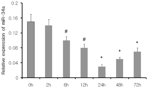
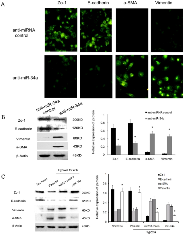
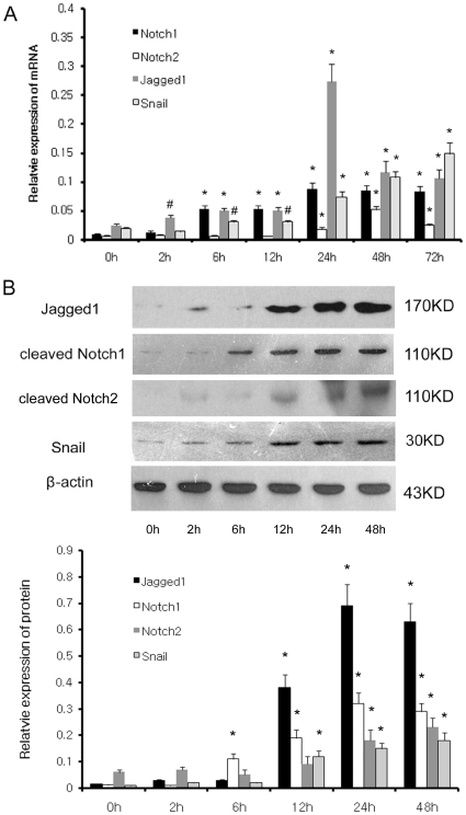
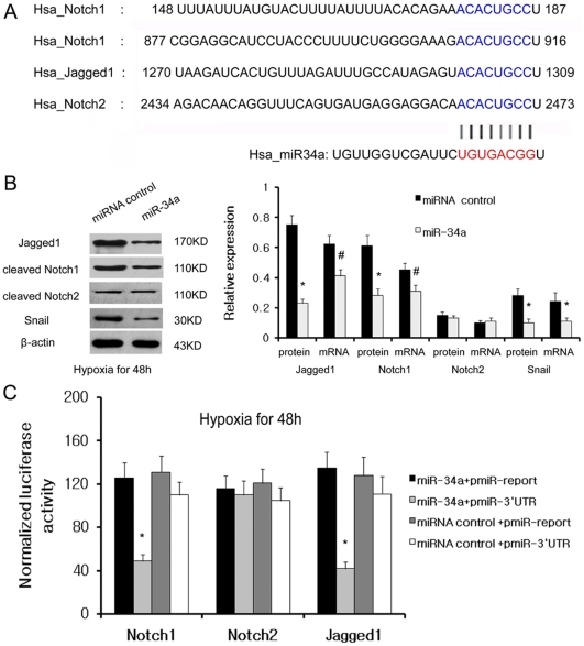
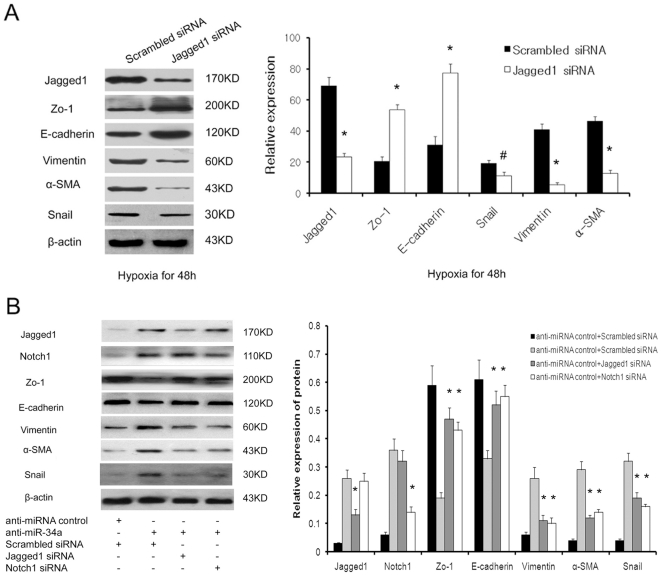
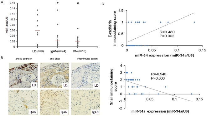
Methodology/principal findings: miRNA profiling was performed for the identification of differentially expressed miRNAs in HK-2 cells under normal and low oxygen, and the results were then verified by quantitative real time RT-PCR (qRT-PCR). The function of miRNAs in hypoxia-induced renal tubular cell EMT was assessed by the transfection of specific miRNA inhibitors and mimics. Luciferase reporter gene assays and western blot analysis were performed to validate the target genes of miR-34a. siRNA against Jagged1 was designed to investigate the role of the miR-34a-Notch pathway in hypoxia induced renal tubular cell EMT. miRNA-34a was identified as being downregulated in hypoxic renal tubular epithelial cells. Inhibition of miR-34a expression in HK-2 cells, which highly express endogenous miR-34a, promoted a mesenchymal phenotype accompanied by reduced expression of the epithelial marker Z0-1, E-cadherin and increased expression of the mesenchymal markers α-SMA and vimentin. Conversely, miR-34a mimics effectively prevented hypoxia-induced EMT. Transfection of miRNA-34a in HK-2 cells under hypoxia abolished hypoxia-induced expression of Notch1 and Jagged1 as well as Notch downstream signals, such as snail. Western blot analysis and luciferase reporter gene assays showed direct evidence for miR-34a targeting Notch1 and Jagged1. siRNAs against Jagged1 or Notch1 effectively prevented miR-34a inhibitor-induced tubular epithelial cell EMT.

Conclusions/significance: Our study provides evidence that the hypoxia-induced decrease of miR-34a expression could promote EMT in renal tubular epithelial cells by directly targeting Notch1 and Jagged1, and subsequently, Notch downstream signaling.


Conflict of interest statement

Competing Interests: The authors have declared that no competing interests exist.

Figures

Figure 1
Figure 1. Hypoxia induces downregulation of miR-34a expression in HK2 cells.
qRT-PCR analysis of miR-34a expression after 2, 6, 12, 24, 48 and 72 h of hypoxia and the relative amount of miRNA was normalized to U6 snRNA. Triplicate assays were performed for each RNA sample. #p<0.05 and *p<0.01 compared with normoxic controls.
Figure 2
Figure 2. miR-34a mediates hypoxia-induced EMT in HK-2 cells.
(A) Immunofluorescence analysis of Zo-1, E-cadherin, α-SMA and vimentin expression in anti-miR-34a and anti-miRNA control transfected HK2 cells. (B) Western blot analysis of Zo-1, E-cadherin, α-SMA and vimentin expression in anti-miR-34a and anti-miRNA control transfected HK2 cells. A representative blot from three independent experiments is shown (left). The histogram shows the average volume density normalized to the loading control, β-actin (right). (C) Western blot analysis of Zo-1, E-cadherin, α-SMA and vimentin expression in parental cells, miR-34a and miRNA control transfected cells after 48 hours under hypoxic conditions. A representative blot from three independent experiments is shown (left). The histogram shows the average volume density normalized to the loading control, β-actin (right). *p<0.01 compared with the parental cells and control cells.
Figure 3
Figure 3. Hypoxia induces Notch signal protein and mRNA expression in HK2 cells.
(A) qRT-PCR analysis of Notch1,Notch2, Jagged1, Snail in HK-2 cells after 2 h, 6 h, 12 h, 24 h, 48 h and 72 h of hypoxia and the relative amount of mRNA was normalized to β-actin. (B) Western blot analysis of Notch1, Notch2, Jagged1, Snail in HK-2 cells after 2 h, 6 h, 12 h, 24 h and 48 h of hypoxia. A representative blot from three independent experiments is shown (upper). The histogram shows the average volume density corrected for the loading control, β-actin (lower). #p<0.05 and *p<0.01 compared with normoxic controls.
Figure 4
Figure 4. Notch1 and Jagged1 are directly regulated by miR-34a.
(A) Putative binding sites of miR-34a in Notch1, Notch2 and Jagge1 3′UTR are shown with color letters. (B) Jagged1, Notch1, Notch2, Snail protein level and mRNA level were respectively detected by Western blotting and qRT-PCR 48 h after transfection with miR-34a precursor or precursor control in HK2 cells under low oxygen. A representative blot from three independent experiments is shown (left). The histogram shows the average volume density normalized to the β-actin (right). (C) Dual luciferase assay was performed in HK2 cells under hypoxia co-transfected with luciferase construct (pMIR-Norch1-3′UTR, pMIR-Norch2-3′UTR, pMIR-Jagged1-3′UTR or pMIR-report control) and miR-34a precursors or precursor control. Firefly luciferase activity was normalized to Renilla luciferase activity for each sample. The results shown represent the mean ± SD from 3 independent experiments. #p<0.05 and *p<0.01 compared with empty vector or miRNA precursor control.
Figure 5
Figure 5. Jagged1 or Notch1 siRNA prevents miR-34a inhibitor-induced EMT via suppression Notch signal.
(A) Jagged1 siRNA prevents hypoxia-induced HK2 cells EMT via suppression Notch signal. Western blots analysis of Zo-1, E-cadherin, α-SMA, vimentin and Snail expression in Jagged1-specific or scrambled siRNA transfected cells after 48 hours under hypoxic conditions. A representative blot from three independent experiments is shown (left). The histogram shows the average volume density normalized to the loading control, β-actin (right). (B) Jagged1 or Notch1 siRNA prevents miR-34a inhibitor-induced HK2 cells EMT via suppression Notch signal. Western blots analysis of Zo-1, E-cadherin, α-SMA, vimentin and Snail expression in Jagged1-specific or scrambled siRNA and miR-34a inhibitor cotransfected HK2 cells. A representative blot from three independent experiments is shown (left). The histogram shows the average volume density normalized to the loading control, β-actin (right). #p<0.05 and *p<0.01 compared with the parental cells and control cells.
Figure 6
Figure 6. Expression of miR-34a in renal tissue from patients with CKD.
(A) Expression analysis of miR-34a in renal tissue from patients with DN, IgA nephropathy (IgAN) by real-time PCR. Shown are relative expression values normalized to U6. Pretransplant biopsies from living donor kidneys (LD) were used as control. *P<0.01. (B) Expression of E-cadherin and Snail in renal tissue from living donor (LD) kidneys and patients with IgA nephropathy (IgAN) by IHC (original magnification, 400×). (C) Scatter plot with fitted values intervals for tubular expression of miR-34a and E-cadherin (upper) or Snail (lower).

Similar articles

Cited by

References

    1. Winter J, Jung S, Keller S, Gregory RI, Diederichs S. Many roads to maturity: microRNA biogenesis pathways and their regulation. Nat Cell Biol. 2009;11:228–234. - PubMed
    1. Ambros V. MicroRNA pathways in flies and worms: Growth, death, fat, stress, and timing. Cell. 2003;113:673–676. - PubMed
    1. Liu Y. Epithelial to mesenchymal transition in renal fibrogenesis: Pathologic significance, molecular mechanism, and therapeutic intervention. J Am Soc Nephrol. 2004;15:1–12. - PubMed
    1. Aigner K, Dampier B, Descovich L, Mikula M, Sultan A, et al. The transcription factor ZEB1 (deltaEF1) promotes tumour cell dedifferentiation by repressing master regulators of epithelial polarity. Oncogene. 2007;26:6979–6988. - PMC - PubMed
    1. Vandewalle C, Comijn J, De Craene B, Vermasse P, Bruyneel E, et al. SIP1/ZEB2 induces EMT by repressing genes of different epithelial cell-cell junctions. Nucleic Acids Res. 2005;33:6566–6578. - PMC - PubMed

Publication types

MeSH terms