Role of position 627 of PB2 and the multibasic cleavage site of the hemagglutinin in the virulence of H5N1 avian influenza virus in chickens and ducks
- PMID: 22363523
- PMCID: PMC3283584
- DOI: 10.1371/journal.pone.0030960
Role of position 627 of PB2 and the multibasic cleavage site of the hemagglutinin in the virulence of H5N1 avian influenza virus in chickens and ducks
Abstract
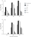
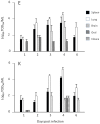
Highly pathogenic H5N1 avian influenza viruses have caused major disease outbreaks in domestic and free-living birds with transmission to humans resulting in 59% mortality amongst 564 cases. The mutation of the amino acid at position 627 of the viral polymerase basic-2 protein (PB2) from glutamic acid (E) in avian isolates to lysine (K) in human isolates is frequently found, but it is not known if this change affects the fitness and pathogenicity of the virus in birds. We show here that horizontal transmission of A/Vietnam/1203/2004 H5N1 (VN/1203) virus in chickens and ducks was not affected by the change of K to E at PB2-627. All chickens died between 21 to 48 hours post infection (pi), while 70% of the ducks survived infection. Virus replication was detected in chickens within 12 hours pi and reached peak titers in spleen, lung and brain between 18 to 24 hours for both viruses. Viral antigen in chickens was predominantly in the endothelium, while in ducks it was present in multiple cell types, including neurons, myocardium, skeletal muscle and connective tissues. Virus replicated to a high titer in chicken thrombocytes and caused upregulation of TLR3 and several cell adhesion molecules, which may explain the rapid virus dissemination and location of viral antigen in endothelium. Virus replication in ducks reached peak values between 2 and 4 days pi in spleen, lung and brain tissues and in contrast to infection in chickens, thrombocytes were not involved. In addition, infection of chickens with low pathogenic VN/1203 caused neuropathology, with E at position PB2-627 causing significantly higher infection rates than K, indicating that it enhances virulence in chickens.
Conflict of interest statement
Figures


References
-
- Li KS, Guan Y, Wang J, Smith GJ, Xu KM, et al. Genesis of a highly pathogenic and potentially pandemic H5N1 influenza virus in eastern Asia. Nature. 2004;430:209–213. - PubMed
-
- WHO. Cumulative number of confirmed human cases of avian influenza A (H5N1) reported to WHO. 2012. http://www.who.int/influenza/human_animal_interface/H5N1_cumulative_tabl..., accessed January 16, 2012.
Publication types
MeSH terms
Substances
LinkOut - more resources
Full Text Sources
Medical
Research Materials
Miscellaneous

