

The Aspergillus nidulans kinesin-3 tail is necessary and sufficient to recognize modified microtubules
- PMID: 22363525
- PMCID: PMC3282709
- DOI: 10.1371/journal.pone.0030976
The Aspergillus nidulans kinesin-3 tail is necessary and sufficient to recognize modified microtubules
Abstract
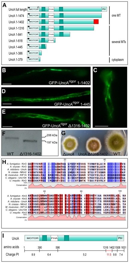
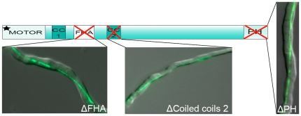
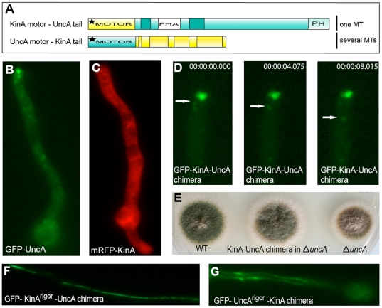
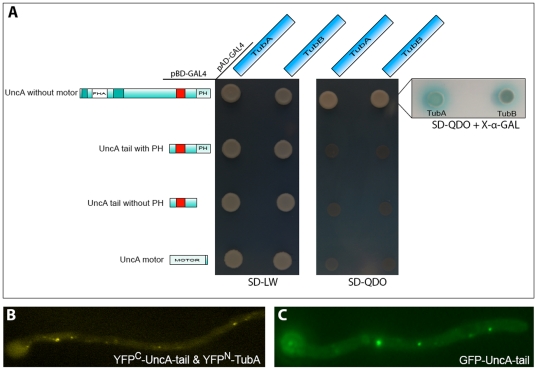
Posttranslational microtubule modifications (PTMs) are numerous; however, the biochemical and cell biological roles of those modifications remain mostly an enigma. The Aspergillus nidulans kinesin-3 UncA uses preferably modified microtubules (MTs) as tracks for vesicle transportation. Here, we show that a positively charged region in the tail of UncA (amino acids 1316 to 1402) is necessary for the recognition of modified MTs. Chimeric proteins composed of the kinesin-1 motor domain and the UncA tail displayed the same specificity as UncA, suggesting that the UncA tail is sufficient to establish specificity. Interaction between the UncA tail and alpha-tubulin was shown using a yeast two-hybrid assay and in A. nidulans by bimolecular fluorescence complementation. This is the first demonstration of how a kinesin-3 motor protein distinguishes among different MT populations in fungal cells, and how specificity determination depends on the tail rather than the motor domain, as has been demonstrated for kinesin 1 in neuronal cells.
Conflict of interest statement
Figures
Similar articles
-
The Aspergillus nidulans kinesin-3 UncA motor moves vesicles along a subpopulation of microtubules.Mol Biol Cell. 2009 Jan;20(2):673-84. doi: 10.1091/mbc.e08-07-0685. Epub 2008 Nov 26. Mol Biol Cell. 2009. PMID: 19037104 Free PMC article.
-
Genetic evidence for a microtubule-destabilizing effect of conventional kinesin and analysis of its consequences for the control of nuclear distribution in Aspergillus nidulans.Mol Microbiol. 2001 Oct;42(1):121-32. doi: 10.1046/j.1365-2958.2001.02609.x. Mol Microbiol. 2001. PMID: 11679072
-
The mitotic kinesin-14 KlpA contains a context-dependent directionality switch.Nat Commun. 2017 Jan 4;8:13999. doi: 10.1038/ncomms13999. Nat Commun. 2017. PMID: 28051135 Free PMC article.
-
Kinesins and microtubules: their structures and motor mechanisms.Curr Opin Cell Biol. 2000 Feb;12(1):35-41. doi: 10.1016/s0955-0674(99)00054-x. Curr Opin Cell Biol. 2000. PMID: 10679355 Review.
-
Emerging Insights into the Function of Kinesin-8 Proteins in Microtubule Length Regulation.Biomolecules. 2018 Dec 20;9(1):1. doi: 10.3390/biom9010001. Biomolecules. 2018. PMID: 30577528 Free PMC article. Review.
Cited by
-
Neurospora crassa NKIN2, a kinesin-3 motor, transports early endosomes and is required for polarized growth.Eukaryot Cell. 2013 Jul;12(7):1020-32. doi: 10.1128/EC.00081-13. Epub 2013 May 17. Eukaryot Cell. 2013. PMID: 23687116 Free PMC article.
-
Microtubule-based transport in filamentous fungi.Curr Opin Microbiol. 2012 Dec;15(6):637-45. doi: 10.1016/j.mib.2012.10.003. Epub 2012 Nov 2. Curr Opin Microbiol. 2012. PMID: 23127389 Free PMC article. Review.
-
Libraries for two-hybrid screening of yeast and hyphal growth forms in Zymoseptoria tritici.Fungal Genet Biol. 2015 Jun;79:94-101. doi: 10.1016/j.fgb.2015.03.023. Fungal Genet Biol. 2015. PMID: 26092795 Free PMC article.
-
Transportation of Aspergillus nidulans Class III and V Chitin Synthases to the Hyphal Tips Depends on Conventional Kinesin.PLoS One. 2015 May 8;10(5):e0125937. doi: 10.1371/journal.pone.0125937. eCollection 2015. PLoS One. 2015. PMID: 25955346 Free PMC article.
-
F-box protein RcyA controls turnover of the kinesin-7 motor KipA in Aspergillus nidulans.Eukaryot Cell. 2014 Aug;13(8):1085-94. doi: 10.1128/EC.00042-14. Epub 2014 Jun 20. Eukaryot Cell. 2014. PMID: 24951440 Free PMC article.
References
-
- Westermann S, Weber K. Post-translational modifications regulate microtubule function. Nat Rev Mol Cell Biol. 2003;4:938–947. - PubMed
-
- Konishi Y, Setou M. Tubulin tyrosination navigates the kinesin-1 motor domain to axons. Nature Neurosci. 2009;12:559–567. - PubMed
-
- Reed NA, Dawen C, Blasius TL, Jih GT, Meyhofer E, et al. Microtubule acetylation promotes kinesin-1 binding and transport. Curr Biol. 2006;16:2166–2172. - PubMed
-
- Dunn S, Morrison EE, Liverpool TB, Molina-Paris C, Cross RA, et al. Differential trafficking of Kif5c on tyrosinated and detryosinated microtubules in live cells. J Cell Sci. 2007;121:1085–1095. - PubMed
-
- Oakley BR. Tubulins in Aspergillus nidulans. Fungal Genet Biol. 2004;41:420–427. - PubMed
Publication types
MeSH terms
Substances
LinkOut - more resources
Full Text Sources
Miscellaneous
