. 2012;7(2):e31056.
doi: 10.1371/journal.pone.0031056. Epub 2012 Feb 17.

Activation of the AMP-activated protein kinase (AMPK) by nitrated lipids in endothelial cells

Affiliations

Activation of the AMP-activated protein kinase (AMPK) by nitrated lipids in endothelial cells

Yong Wu et al. PLoS One. 2012.

Retraction in

Abstract

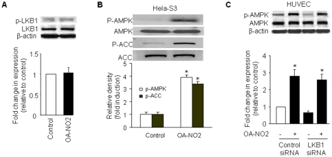
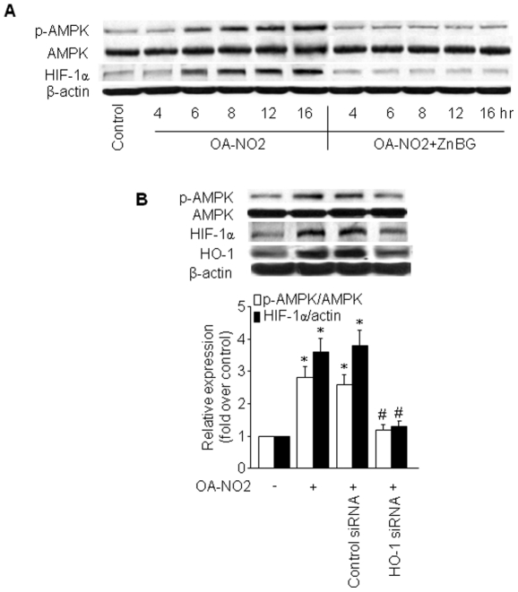
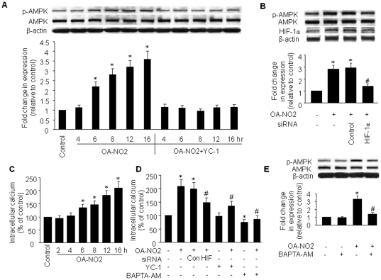
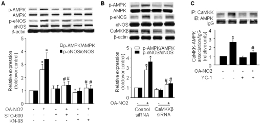
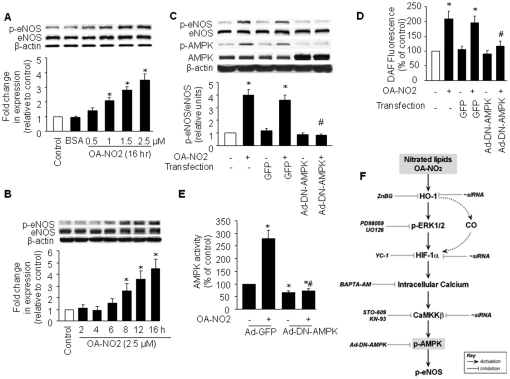
The AMP-activated protein kinase (AMPK) is an important regulator of endothelial metabolic and functional homeostasis. Here, we examined the regulation of AMPK by nitrated oleic acid (OA-NO(2)) and investigated the implications in endothelial function. Treatment of bovine aortic endothelial cells (BAECs) with OA-NO(2) induced a significant increase in both AMPK-Thr172 phosphorylation and AMPK activity as well as upregulation of heme oxygenase (HO)-1 and hypoxia-inducible factor (HIF)-1α. Pharmacologic inhibition or genetic ablation of HO-1 or HIF-1α abolished OA-NO(2)-induced AMPK phosphorylation. OA-NO(2) induced a dramatic increase in extracellular signal-regulated kinase (ERK)1/2 phosphorylation that was abrogated by the HO-1 inhibitor, zinc deuteroporphyrin IX 2,4-bis-ethylene glycol (ZnBG). Inhibition of ERK1/2 using UO126 or PD98059 reduced but did not abolish OA-NO(2)-induced HIF-1α upregulation, suggesting that OA-NO(2)/HO-1-initiated HIF-1α induction is partially dependent on ERK1/2 activity. In addition, OA-NO(2) enhanced endothelial intracellular Ca(2+), an effect that was inhibited by the HIF-1α inhibitor, YC-1, and by HIF-1α siRNA. These results implicate the involvement of HIF-1α. Experiments using the Ca(2+)/calmodulin-dependent protein kinase kinase (CaMKK) inhibitor STO-609, the selective CaMKII inhibitor KN-93, and an isoform-specific siRNA demonstrated that OA-NO(2)-induced AMPK phosphorylation was dependent on CaMKKβ. Together, these results demonstrate that OA-NO(2) activates AMPK in endothelial cells via an HO-1-dependent mechanism that increases HIF-1α protein expression and Ca(2+)/CaMKKβ activation.


Conflict of interest statement

Competing Interests: The authors have declared that no competing interests exist.

Figures

Figure 1
Figure 1. Induction of HO-1 protein and AMPK activation by OA-NO2.
A) BAECs were incubated with OA-NO2 at the indicated concentrations or with BSA (vehicle) for 16 h, and western blot analysis was performed as described in Materials and Methods to detect HO-1 protein expression and AMPK phosphorylation at Thr172. The blot is representative of those obtained from three separate experiments. Corresponding densitometric analyses of phosphorylated AMPK and ACC are shown. *p<0.05 vs. control. B) BAECs were incubated with 2.5 µM OA-NO2 for the indicated times, and western blotting was performed as above. The blot is representative of three blots obtained from three separate experiments. *p<0.05 vs. corresponding control. C) Confluent BAECs were exposed to vehicle or OA-NO2 (2.5 µM) for 16 h. AMPKα was immunoprecipitated from cell lysates (1 mg) with a specific antibody. AMPK activity was assayed by 32P-ATP incorporation into the SAMS peptide. *p<0.05 vs. control. D) BAECs were incubated with the indicated concentrations of OA for 16 h. Western blotting was performed as described in Materials and Methods. E) BAECs were infected with Ad-DN-AMPK (MOI = 50) or Ad-GFP (control). Infected and non-infected cells were treated with 2.5 µM OA-NO2 for 16 h. AICAR and metformin were used as positive controls. The blot is representative of three blots obtained from three separate experiments.
Figure 2
Figure 2. Activation of AMPK by OA-NO2 does not require LKB1.
A) Phosphorylation of LKB1 Ser428 was not affected by OA-NO2 in BAECs. Confluent BAECs were exposed to 2.5 µM OA-NO2 for 16 h, and phosphorylated LKB1-Ser428 was detected by a phospho-specific antibody in western blots. The blot is a representative of three blots obtained from three independent experiments. Lower panels: summary data (n = 3). B) LKB1 is not required for AMPK activation by OA-NO2. Confluent LKB1-deficient Hela-S3 cells were exposed to 2.5 µM OA-NO2 for 16 h, and then AMPK and ACC phosphorylation were assayed as described in Materials and Methods. The blot is representative of three blots obtained from three independent experiments. Lower panels: summary data (*p<0.05 vs. control; n = 3). C) LKB1 siRNA did not abolish OA-NO2-stimulated AMPK activation in HUVECs. HUVECs were incubated with LKB1-specific siRNA or control siRNA for 48 h and then treated with OA-NO2 or vehicle for 16 h. After treatment, cell lysates were analyzed for LKB1 protein levels and AMPK phosphorylation at Thr172. Lower panels: summary data (*p<0.05 vs. control; n = 3).
Figure 3
Figure 3. OA-NO2-induced HIF-1α is dependent on HO-1.
A) HUVECs were treated with 1 µM ZnBG for 30 min followed by incubation with OA-NO2 for the indicated times. AMPK protein and phosphorylation levels and HIF-1α protein expression were assayed as described in Materials and Methods. The blot is representative of three blots obtained from three independent experiments. B) HUVECs were incubated with HO-1-specific siRNA or control siRNA for 48 h and then treated with OA-NO2 or vehicle for 16 h. After treatment, cell lysates were analyzed for HIF-1α and HO-1 protein levels and AMPK phosphorylation at Thr172. Lower panels: summary data (*p<0.05 vs. control; # p<0.05 vs. OA-NO2 group; n = 3).
Figure 4
Figure 4. OA-NO2-induced HIF-1α is partially dependent on ERK1/2.
A) BAECs were incubated with the indicated concentrations of OA-NO2 for 16 h. Protein expression and phosphorylation of p38 MAPK and ERK1/2 were assayed as described in Materials and Methods. The blot is representative of three blots obtained from three independent experiments. B) BAECs were treated with ZnBG (1 µM) for 30 min followed by incubation with OA-NO2 for 16 h. ERK1/2 phosphorylation levels were monitored by immunoblot analysis, and band density was normalized to total ERK1/2 levels (lower panel). The blot is representative of three blots obtained from three independent experiments. *p<0.05 vs. control; # p<0.05 vs. OA-NO2 group. C) BAECs were treated with OA-NO2 alone or with the ERK1/2 signaling inhibitors, UO126 (10 µM) or PD98059 (50 µM), 1 h before the addition of OA-NO2. HIF-1α protein levels were monitored by immunoblot analysis, and band density was normalized to β-actin levels (lower panel). The blot is representative of three blots obtained from three independent experiments. *p<0.05 vs. control; # p<0.05 vs. OA-NO2 group.
Figure 5
Figure 5. HIF-1α mediates OA-NO2-induced intracellular Ca2+ accumulation and AMPK activation.
A) BAECs were treated with YC-1 (30 µM) for 30 min followed by incubation with OA-NO2 for the indicated times. AMPK phosphorylation and protein levels were assayed as described in Materials and Methods. The blot is representative of three blots obtained from three independent experiments. B) BAECs were incubated with HIF-1α-specific siRNA or control siRNA for 48 h and then treated with OA-NO2 or vehicle for 16 h. After treatment, cell lysates were analyzed for HIF-1α and AMPK protein levels and AMPK phosphorylation at Thr172. Lower panels: summary data (*p<0.05 vs. control; # p<0.05 vs. OA-NO2 group; n = 3). C) BAECs were incubated with OA-NO2 (2.5 µM) for the indicated times. Intracellular Ca2+ was measured with Fluo-4 fluorescent dye as described in Materials and Methods. *p<0.01 vs. control (n = 4). D) BAECs were pre-treated with YC-1 (30 µM) or BAPT-AM (25 µM) for 30 min or HIF-1α siRNA or control siRNA for 48 h followed by incubation with OA-NO2 for 16 h. After treatment, intracellular Ca2+ was measured in intact cells using a Fluo-4 NW kit. *p<0.05 vs. control; # p<0.05 vs. OA-NO2 group (n = 3). E) BAECs were pre-loaded with 25 µM BAPT-AM for 30 min prior to incubation with 2.5 µM OA-NO2 for 16 h. AMPK protein levels and phosphorylation at Thr172 were detected as described above. Representative blots (top) and densitometric analyses (bottom) are shown. Values are means ± SD from three independent measurements. *p<0.05 vs. control; # p<0.05 vs. OA-NO2 group.
Figure 6
Figure 6. CaMKK is the upstream AMPKK that mediates OA-NO2-induced AMPK activation.
A) HUVECs were treated with STO-609 (1 µM) or KN-93 (3 µM) for 1 h followed by incubation with OA-NO2 for 16 h. AMPK and eNOS phosphorylation and protein expression were assayed as described in Materials and Methods. The blot is representative of three blots obtained from three independent experiments. Lower panels: summary data (*p<0.05 vs. control; # p<0.05 vs. OA-NO2 group; n = 3). B) HUVECs were incubated with CaMKKβ-specific siRNA or control siRNA for 48 h and then treated with OA-NO2 for 16 h. After treatment, cell lysates were analyzed for AMPK and eNOS phosphorylation and protein levels. Lower panels: summary data (*p<0.05 vs. control; # p<0.05 vs. OA-NO2 group; n = 3). C) HUVECs were pre-treated with YC-1 (30 µM) for 30 min followed by incubation with OA-NO2 for 16 h. An immunoblot of AMPK precipitated with an anti-CaMKK antibody is shown. The blot is representative of three blots obtained from three independent experiments. Lower panels: summary data (*p<0.05 vs. control; # p<0.05 vs. OA-NO2 group; n = 3).
Figure 7
Figure 7. AMPK mediates OA-NO2-induced eNOS phosphorylation and NO production in BAECs.
BAECs were treated with (A) different concentrations of OA-NO2 for 16 h or (B) 2.5 µmol/L OA-NO2 for the indicated times. Lysates were analyzed by western blot for the indicated proteins. The blot is representative of three blots obtained from three separate experiments. C) Western blot of phosphorylated AMPK and eNOS in OA-NO2-stimulated BAECs infected with Ad-DN-AMPK (MOI = 50). Non-infected cells or cells infected with Ad-GFP served as controls. For A–C, the corresponding densitometric analyses are shown. *p<0.05 vs. control; # p<0.05 vs. GFP with OA-NO2-treated group. D) NO release by OA-NO2-stimulated BAECs infected with Ad-DN-AMPK (MOI = 50) or Ad-GFP (control). *p<0.05 vs. non-transfected, no OA-NO2 group; # p<0.05 vs. OA-NO2-treated, Ad-GFP group. E) AMPK activity corresponding to C and D above. *p<0.05 vs. no OA-NO2 treatment, Ad-GFP group; # p<0.05 vs. OA-NO2-treated, Ad-GFP group. F) The proposed signaling pathway involved in AMPK/eNOS activation in response to OA-NO2 treatment in endothelial cells.

Similar articles

Cited by

References

    1. Ischiropoulos H, Beckman JS. Oxidative stress and nitration in neurodegeneration: cause, effect, or association? J Clin Invest. 2003;111:163–169. - PMC - PubMed
    1. Zou MH, Cohen R, Ullrich V. Peroxynitrite and vascular endothelial dysfunction in diabetes mellitus. Endothelium. 2004;11:89–97. - PubMed
    1. Radi R. Nitric oxide, oxidants, and protein tyrosine nitration. Proc Natl Acad Sci U S A. 2004;101:4003–4008. - PMC - PubMed
    1. Baker PR, Lin Y, Schopfer FJ, Woodcock SR, Groeger AL, et al. Fatty acid transduction of nitric oxide signaling: multiple nitrated unsaturated fatty acid derivatives exist in human blood and urine and serve as endogenous peroxisome proliferator-activated receptor ligands. J Biol Chem. 2005;280:42464–42475. - PMC - PubMed
    1. Villacorta L, Zhang J, Garcia-Barrio MT, Chen XL, Freeman BA, et al. Nitro-linoleic acid inhibits vascular smooth muscle cell proliferation via the Keap1/Nrf2 signaling pathway. Am J Physiol Heart Circ Physiol. 2007;293:H770–776. - PMC - PubMed

Publication types

MeSH terms

Substances