. 2012;7(2):e31140.
doi: 10.1371/journal.pone.0031140. Epub 2012 Feb 17.

Brainstem respiratory oscillators develop independently of neuronal migration defects in the Wnt/PCP mouse mutant looptail

Affiliations

Brainstem respiratory oscillators develop independently of neuronal migration defects in the Wnt/PCP mouse mutant looptail

Muriel Thoby-Brisson et al. PLoS One. 2012.

Abstract

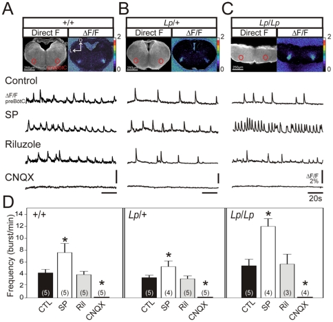
The proper development and maturation of neuronal circuits require precise migration of component neurons from their birthplace (germinal zone) to their final positions. Little is known about the effects of aberrant neuronal position on the functioning of organized neuronal groups, especially in mammals. Here, we investigated the formation and properties of brainstem respiratory neurons in looptail (Lp) mutant mice in which facial motor neurons closely apposed to some respiratory neurons fail to migrate due to loss of function of the Wnt/Planar Cell Polarity (PCP) protein Vangl2. Using calcium imaging and immunostaining on embryonic hindbrain preparations, we found that respiratory neurons constituting the embryonic parafacial oscillator (e-pF) settled at the ventral surface of the medulla in Vangl2(Lp/+) and Vangl2(Lp/Lp) embryos despite the failure of tangential migration of its normally adjacent facial motor nucleus. Anatomically, the e-pF neurons were displaced medially in Lp/+ embryos and rostro-medially Lp/Lp embryos. Pharmacological treatments showed that the e-pF oscillator exhibited characteristic network properties in both Lp/+ and Lp/Lp embryos. Furthermore, using hindbrain slices, we found that the other respiratory oscillator, the preBötzinger complex, was also anatomically and functionally established in Lp mutants. Importantly, the displaced e-pF oscillator established functional connections with the preBötC oscillator in Lp/+ mutants. Our data highlight the robustness of the developmental processes that assemble the neuronal networks mediating an essential physiological function.


Conflict of interest statement

Competing Interests: The authors have declared that no competing interests exist.

Figures

Figure 1
Figure 1. Location of the facial motor nucleus and Phox2b-positive cells in the hindbrain of Lp mutant embryos.
A–C: In situ hybridization of Egr2 (black) and Hoxb1 (red) in E8.5 whole hindbrain preparations (lateral view) obtained from a +/+ (A), Lp/+ (B) and Lp/Lp (C) embryos. Wt (+/+) embryo processed for Egr2 in situ only. Expression of Egr2 in rhombomere 3 (r3) and r5, and of Hoxb1 in r4 are unaffected in Lp/+ and Lp/Lp embryos. D–F: In situ hybridization of Tbx20 expression in E14.5 whole hindbrain preparations (ventral view) obtained from +/+ (D), Lp/+ (E) and Lp/Lp (F) embryos. Facial motoneurons failed to migrate properly both in Lp/+ and Lp/Lp embryos. The black squares indicate the regions illustrated in G, H and I. G–O: Anti-Phox2b (red) and anti-Islet1,2 (green) immunofluorescence on whole hindbrain preparations (G–I, ventral view) and transverse sections (J–O) through E15.5 hindbrains of +/+ (G, J, M), Lp/+ (H, K, N) and Lp/Lp (I, L, O) embryos. White arrowheads in J and K point to Phox2b-positive cells located ventral to the facial nucleus. Numbered dashed lines in G–I refer for each genotype to the axial level corresponding to the sections illustrated in J–R. P–R: Anti-NK1R (red) and anti-Islet1,2 (green) immunofluorescence on transverse sections through E15.5 hindbrain preparations obtained at level 1 for a +/+ embryo (P), and at level 2 for Lp/+ (Q) and Lp/Lp (R) embryos. Slices illustrated in M and P, N and Q, O and R, correspond to adjacent slices obtained from the same animal, thus showing possible co-expression of several markers in the same neuronal population. Immunostainings indicate the presence of Phox2b-positive or NK1R-positive cell groups present at the ventral surface of the hindbrain for all genotypes. D: dorsal, L: lateral, R: rostral.
Figure 2
Figure 2. Rostro-caudal distribution of facial motor neurons and respiratory neurons in the hindbrain of wild-type and Lp mutant embryos.
Half slice drawings were obtained from images of brainstem slices labeled with anti-NK1R and Islet1,2 antibodies (dorsal up). The red dots indicate individual e-pF neurons, the gray areas indicate the FMN and the blue ovals indicate the preBötC. The rostro-caudal extents of the preBötC (blue), the e-pF (red) and the FMN (gray) are indicated by vertical colored bars. The numbers on the left of each drawing indicate the rostro-caudal position (in µm) of the slice relative to the preBötC position, which is defined as zero (see the full scale on the left of +/+). Note that the e-pF is more rostrally and loosely distributed in the Lp/Lp preparation compared to the +/+ and Lp/+ preparations. Similar distributions were observed in a second preparation for each genotype. XIIn: hypoglossal nucleus, nA: nucleus ambiguus, FMN: facial motor nucleus.
Figure 3
Figure 3. Altered distribution of active cells at the pial surface of Lp mutant hindbrain preparations.
A–C: Ventral view of whole hindbrain preparations from E15.5 +/+ (A), Lp/+ (B) and Lp/Lp (C) embryos loaded with Calcium Green 1-AM and observed in direct fluorescence. Yellow ovals indicate the position of the facial motor nucleus (FMN) that is clearly visible in direct light (see right side of preparations in A and B). D–F: Maps for rhythmic active cells (red circles) detected at a high magnification for corresponding genotypes in the region delimited by the white rectangles in A–C. Active cells located at the ventral surface of the preparations are found for all genotypes. G–I: Histograms of the rostro-caudal distribution of active cells relative to the constant preBötC position for 2 wild-type (G), 3 Lp/+ (H), and 3 Lp/Lp (I) embryos. The red arrows indicate the rostral and the caudal extremity of the FMN. The distribution of active cells shows a significant rostral displacement in Lp/Lp embryos. J–L: Calcium transients illustrated as relative fluorescent changes (ΔF/F) recorded in the region encompassing the active cells (delimited by the blue rectangles in A–C). Relative fluorescent changes intensity are color-coded, white corresponding to the strongest activity (see the color scale at the bottom). The traces below show the spontaneous calcium changes recorded over time in the entire active region for each genotype. R: rostral.
Figure 4
Figure 4. Active cells form a functional e-pF oscillator in Lp mutant embryos.
A: Ventral view of the hindbrain over the area encompassing the FMN and the active region. Preparations were obtained from an E15.5 +/+ embryo, loaded with calcium Green 1-AM and observed in direct fluorescence (left panel) or as relative changes in fluorescence (ΔF/F, right panel). The yellow oval indicates the position of the FMN, the e-pF network is encircled in red. Traces below indicate calcium transients (ΔF/F) measured in the e-pF region in control conditions (top trace), pH 7.2 (second trace), 10 µM CNQX (third trace), 10 µM Riluzole (fourth trace) and 10−7 M Substance P (SP, fifth trace). B and C: Same legend as in A for a Lp/+ embryo (B) and a Lp/Lp embryo (C). D: Graphs representing the mean frequency of calcium transients measured in the e-pF region in different experimental conditions (indicated below and color coded) for +/+ (left part), Lp/+ (middle part) and Lp/Lp (right part). Numbers in brackets indicate the number of preparations tested. Asterisks indicate statistically different means. Frequency is increased in the presence of CNQX, SP and in pH 7.2 for all genotypes, while riluzole blocks the rhythmic activity in the active region. The active network found in the mutant shares pharmacological characteristics of the e-pF recorded in the wild type littermates. L: lateral, R: rostral.
Figure 5
Figure 5. Bilateral e-pF oscillators exhibit synchronized activity in Lp mutant embryos.
A–C: Left: Ventral view of whole hindbrain preparations from E15.5 +/+ (A), Lp/+ (B) and Lp/Lp (C) embryos loaded with Calcium Green 1-AM and observed in direct fluorescence. The red lines indicate the position of the left (L) and the right (R) e-pF oscillators. The two traces (middle panels) correspond to the calcium transients illustrated as relative fluorescent changes (ΔF/F) recorded simultaneously in the corresponding e-pF oscillators in one preparation. The dashed lines in blue highlight the synchronicity between the activities of both e-pF oscillators. Right: corresponding superimposed left-right e-pF cross-correlograms illustrated for 4 different preparations. Bilateral synchronous activity in the two e-pF oscillators is preserved the Lp/+ and Lp/Lp mutants. R: rostral.
Figure 6
Figure 6. The preBötC oscillator is preserved in Lp/Lp mutants.
Anti-NK1R (red) and anti-Islet1,2 (green) immunofluorescence on hindbrain transverse slices obtained from E16.5 embryos for +/+ (A, A′), Lp/+ (B, B′) and Lp/Lp (C, C′) genotypes at the level of the preBötC area. The panels in A′–C′ are higher magnification views of the preBötC region delimited by white rectangles in A–C, respectively. The preBötC oscillator is defined as the NK1R-positive region located ventrally to the Islet1,2-positive nucleus ambiguous (nA). The preBötC is anatomically preserved in the Lp/Lp mutant. D: dorsal, L: lateral.
Figure 7
Figure 7. Functional analysis of the preBötC oscillator in the Lp mutant.
A: Photomicrograph of a transverse medullary slice through the preBötC obtained from an E16.5 +/+ embryo loaded with calcium green 1-AM observed in direct fluorescence (left panel) and as relative changes in fluorescence (ΔF/F, right panel). The red circles indicate the position of the bilaterally distributed preBötC oscillators. Traces below (ΔF/F preBötC) indicate calcium transients measured in the preBötC region in control conditions (top trace), 10−7 M Substance P (SP, second trace), 10 µM Riluzole (third trace) and 10 µM CNQX (fourth trace). B and C: Same legend as in A for a Lp/+ embryo (B) and a Lp/Lp embryo (C). D: Graph representing the mean frequency of calcium transients measured in the preBötC region in different experimental conditions (indicated below and color coded) for +/+ (left part), Lp/+ (middle part) and Lp/Lp (right part). Numbers in brackets indicate the number of preparations tested. Asterisks indicate statistically different means. Frequency is increased in the presence of SP, unchanged in the presence of riluzole and blocked in the presence of CNQX for all genotypes. Hence, the preBötC oscillator is functionally preserved in Lp/Lp mutants. D: dorsal, L: lateral.
Figure 8
Figure 8. Response to a low pH challenge is unaffected in the Lp/+ mutant.
Integrated phrenic nerve discharge (Int C4) at pH 7.4 (upper trace) and pH 7.2 (bottom traces) for control (A) and Lp/+ (B) embryos at E16.5. C: Quantification of burst frequencies for control (left) and heterozygous (right) embryos at pH 7.4 (white bars) and pH 7.2 (gray bars). Numbers of hindbrain preparations analyzed are indicated on the bars. The motor output of the respiratory network recorded from C4 nerve roots is comparable in Lp/+ and wild type preparations in control conditions, and the response to low pH is preserved in the Lp/+ mutant.
Figure 9
Figure 9. Schematic summary of the relative positions of respiratory oscillators and the FMN in the hindbrain in +/+, Lp/+ and Lp/Lp embryos.
Hindbrain outlines were drawn using photomicrographs of isolated preparations as a template. The dashed blue line indicates the landmark used to align the preparations (position of the caudal limit of migrating pontine neurons). The red dashed lines indicate the rostral and caudal limits of the e-pF oscillator (red ovals). The blue ovals indicate the preBötzinger complex. The position of the facial motor nucleus (FMN) is indicated by the black ovals, shown with a dashed line in the Lp/Lp embryo due to its dorsal position. The e-pF oscillator is slightly displaced rostro-medially in Lp/+ embryos, and significantly displaced rostrally in Lp/Lp embryos relative to +/+ embryos.

Similar articles

Cited by

References

    1. Guerrini R, Parrini E. Neuronal migration disorders. Neurobiol Dis. 2010;38:154–166. - PubMed
    1. Verrotti A, Spalice A, Ursitti F, Papetti L, Mariani R, et al. New trends in neuronal migration disorders. Eur J Paediatr Neurol. 2010;14:1–12. - PubMed
    1. Aicardi J. The place of neuronal migration abnormalities in child neurology. Can J Neurol Sci. 1994;21:185–193. - PubMed
    1. Smith JC, Ellenberger HH, Ballanyi K, Richter DW, Feldman JL. Pre-Botzinger complex: a brainstem region that may generate respiratory rhythm in mammals. Science. 1991;254:726–729. - PMC - PubMed
    1. Janczewski WA, Feldman JL. Distinct rhythm generators for inspiration and expiration in the juvenile rat. J Physiol. 2006;570:407–420. - PMC - PubMed

Publication types

MeSH terms

LinkOut - more resources