. 2012;7(2):e31176.
doi: 10.1371/journal.pone.0031176. Epub 2012 Feb 17.

Establishment of motor neuron-V3 interneuron progenitor domain boundary in ventral spinal cord requires Groucho-mediated transcriptional corepression

Affiliations

Establishment of motor neuron-V3 interneuron progenitor domain boundary in ventral spinal cord requires Groucho-mediated transcriptional corepression

Keith J Todd et al. PLoS One. 2012.

Abstract

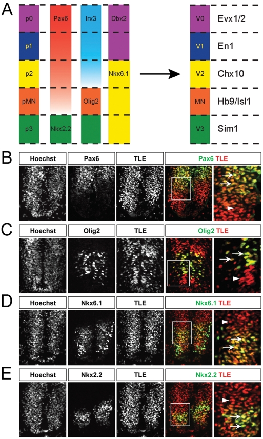
Background: Dorsoventral patterning of the developing spinal cord is important for the correct generation of spinal neuronal types. This process relies in part on cross-repressive interactions between specific transcription factors whose expression is regulated by Sonic hedgehog. Groucho/transducin-like Enhancer of split (TLE) proteins are transcriptional corepressors suggested to be recruited by at least certain Sonic hedgehog-controlled transcription factors to mediate the formation of spatially distinct progenitor domains within the ventral spinal cord. The aim of this study was to characterize the involvement of TLE in mechanisms regulating the establishment of the boundary between the most ventral spinal cord progenitor domains, termed pMN and p3. Because the pMN domain gives rise to somatic motor neurons while the p3 domain generates V3 interneurons, we also examined the involvement of TLE in the acquisition of these neuronal fates.

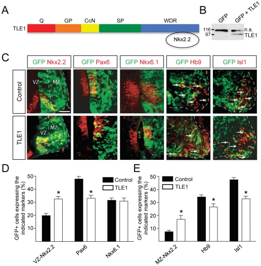
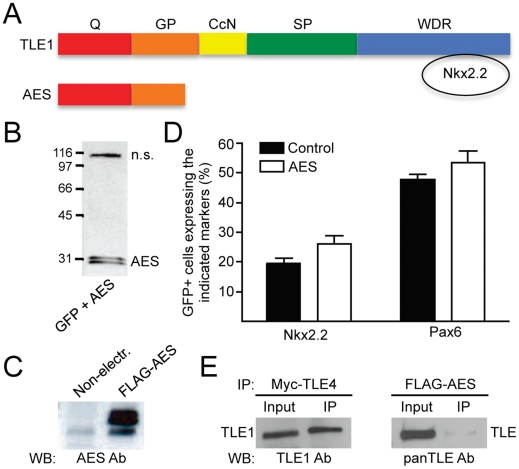
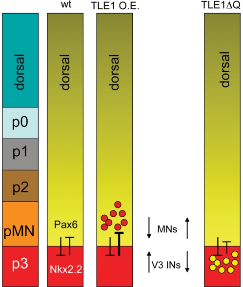
Methodology and principal findings: A combination of in vivo loss- and gain-of-function studies in the developing chick spinal cord was performed to characterize the role of TLE in ventral progenitor domain formation. It is shown here that TLE overexpression causes increased numbers of p3 progenitors and promotes the V3 interneuron fate while suppressing the motor neuron fate. Conversely, dominant-inhibition of TLE increases the numbers of pMN progenitors and postmitotic motor neurons.

Conclusion: Based on these results, we propose that TLE is important to promote the formation of the p3 domain and subsequent generation of V3 interneurons.


Conflict of interest statement

Competing Interests: The authors have declared that no competing interests exist.

Figures

Figure 1
Figure 1. TLE expression in the embryonic chick spinal cord.
(A) Schematic representation of the five progenitor cell (p) domains of the ventral spinal cord, termed p0, p1, p2, pMN and p3 from dorsal to ventral positions, respectively. These domains are defined by the specific expression of combinations of HD and bHLH transcription factors. Refinement and maintenance of these progenitor domains is achieved through cross-repressive interactions between pairs of transcription factors, for example between Pax6 and Nkx2.2 at the pMN/p3 boundary. In turn, each progenitor domain generates different neuronal populations, V0, V1 and V2 INs, somatic MNs and V3 INs, respectively. Like the progenitor domains, separate populations of postmitotic neurons can be defined by the expression of specific transcription factors, such as HB9 and Isl1 in MNs derived from the pMN domain or other factors in other cell types, as indicated in the right-hand column. (B–E) Sections through the spinal cord of HH stage 18 chick embryos were subjected to double-labeling immunohistochemical analysis using a panTLE antibody together with antibodies against the indicated proteins. Panels in the right-hand column show high-magnification views of the boxed areas in the adjacent panels. Arrows point to examples of double-labeled cells. Arrowheads point to examples of cells expressing only TLE. TLE expression was observed in most ventral spinal cord cells, including domains p0–p2 (region of high Pax6 immunoreactivity dorsal to the Olig2+ domain), pMN (region expressing Nkx6.1, Olig2 and low levels of Pax6) and p3 (region expressing Nkx6.1 and Nkx2.2) of the ventral area. Notice in particular how virtually all Nkx2.2+ cells also express TLE.
Figure 2
Figure 2. Effect of TLE1 overexpression in the developing chick ventral spinal cord.
(A) Schematic representation of the TLE domain structure. Notice the Q domain involved in oligomerization and transcriptional repression and the WDR domain important for protein-protein interactions . Nkx2.2 binds to the TLE WDR domain using an Eh-1 motif. (B) Western blotting analysis of lysates from chick embryo spinal cords electroporated with plasmids encoding GFP alone or together with FLAG epitope-tagged TLE1 demonstrating the expression of exogenous TLE1 using anti-FLAG antibody. “n.s.” indicates a non-specific band detected by this antibody. (C) Double-labeling analysis of the expression of GFP and the indicated proteins in embryos electroporated with GFP alone (control) or GFP+TLE1 (TLE1). Nkx2.2+ cells were observed in both the ventricular zone (VZ) and mantle zone (MZ). Arrows in the two right-hand columns point to examples of double-labeled cells coexpressing GFP and either Hb9 or Isl1. (D and E) Quantification of the numbers of electroporated cells (GFP+) expressing Nkx2.2 [in either the VZ (D) or the MZ (E)], Pax6, Nkx6.1, Hb9, or Isl1, as indicated. TLE1 overexpression caused an increase in the number of Nkx2.2+ cells in the VZ, with a concomitant decrease in Pax6+ cells. The number of cells expressing Nkx6.1 was not altered. These changes were associated with an increase in Nkx2.2+ cells in the MZ and a decrease in the number of electroporated cells expressing the MN markers Hb9 and Isl1. Data are expressed as mean ± SEM (*p<0.05). Scale bars = 50 µm.
Figure 3
Figure 3. Lack of effect of AES overexpression on ventral spinal cord Pax6+ and Nkx2.2+ progenitor populations.
(A) Schematic comparison of the structure of AES to that of full-length TLE. AES lacks the WDR domain involved in Nkx2.2 binding but retains the amino-terminal Q domain . (B and C) Western blotting analysis of lysates from chick embryo spinal cords electroporated with plasmids encoding GFP together with FLAG epitope-tagged AES using either anti-FLAG (B) or anti-AES (C) antibodies. (B) “n.s.” indicates a non-specific band detected by the anti-FLAG antibody. (C) Exogenous AES was dramatically overexpressed in electroporated spinal cords. (D) Quantification of the number of GFP+ cells expressing Nkx2.2 or Pax6 in chick embryos electroporated with GFP alone (Control) or together with AES (AES). AES had no significant effect on the number of either Nkx2.2+ or Pax6+ progenitor cells. (E) Coimmunoprecipitation experiments performed using lysates from chick embryo spinal cords electroporated with plasmids encoding either Myc-tagged TLE4 or FLAG-tagged AES, as indicated. TLE4 or AES were immunoprecipitated (IP) using anti-Myc or anti-FLAG antibodies, respectively, followed by Western blotting (WB) analysis of input lysate (10%) and immunoprecipitated material using an anti-TLE1 antibody that does not cross-react with TLE4 or a panTLE antibody that recognizes all full-length TLE proteins because it is directed against the WDR domain . Endogenous TLE1 coimmunoprecipitated efficiently with exogenous TLE4. In contrast, only a modest coimmunoprecipitation of AES with endogenous TLE was detected.
Figure 4
Figure 4. Effect of TLE1ΔQ expression on ventral spinal cord Pax6+ and Nkx2.2+ progenitor populations and neuronal fate acquisition.
(A) Schematic representation of TLE1ΔQ, compared to TLE1 and AES, depicting the lack of the Q domain but retention of the WDR domain in TLE1ΔQ. (B) Coimmunoprecipitation experiments performed using lysates from chick embryo spinal cords electroporated with plasmid encoding FLAG-TLE1ΔQ. Immunoprecipitation (IP) was performed using anti-FLAG antibody, followed by Western blotting (WB) analysis of input lysate (10%) and immunoprecipitated material using a panTLE antibody that recognizes all full-length TLE proteins and also TLE1ΔQ because it is directed against the WDR domain . Endogenous TLE did not coimmunoprecipitate with exogenous TLE1ΔQ. (C) Quantification of the number of GFP+ cells expressing Nkx2.2 [in either the ventricular zone (VZ) or marginal zone (MZ)], Pax6, Hb9, or Isl1 in chick embryos electroporated with GFP alone or together with TLE1 or TLE1ΔQ. Expression of TLE1ΔQ resulted in an increase in the number of Pax6+ progenitor cells as well as Hb9+ and Isl1+ MNs compared to the control conditions. These effects were opposite to the effects of TLE1. See Figure S5 for double-labeling immunohistochemical analysis of electroporated embryos. (D) Quantification of the number of GFP+ cells expressing Pax6 in chick embryo spinal cord electroporated with GFP alone or together with TLE1, TLE1ΔQ, or TLE1 and TLE1ΔQ together, as indicated. Data in (C and D) are expressed as mean ± SEM (*p<0.05; **p<0.01; n.s., not significant).
Figure 5
Figure 5. Schematic summary of the effects of TLE perturbations on Pax6+ and Nkx2.2+ ventral progenitor populations and neuronal fate acquisition.
Pax6 and Nkx2.2 normally repress the expression of each other to establish the pMN/p3 boundary. Overexpression (O.E.) of TLE1 increases the number of Nkx2.2+ progenitor cells and V3 INs at the expense of Pax6+ progenitor cells and somatic MNs. Conversely, exogenous expression of TLE1ΔQ results in an increase in Pax6+ progenitor cells and MNs.

Similar articles

Cited by

References

    1. Briscoe J, Ericson J. Specification of neuronal fates in the ventral neural tube. Curr Opin Neurobiol. 2001;11:43–49. - PubMed
    1. Zhuang B, Sockanathan S. Dorso-ventral patterning: a view from the top. Curr Opin Neurobiol. 2006;16:20–24. - PubMed
    1. Dessaud E, McMahon AP, Briscoe J. Pattern formation in the vertebrate neural tube: a sonic hedgehog morphogen-regulated transcriptional network. Development. 2008;135:2489–2503. - PubMed
    1. Lupo G, Harris WA, Lewis KE. Mechanisms of ventral patterning in the vertebrate nervous system. Nat Rev Neurosci. 2006;7:103–114. - PubMed
    1. Lu QR, Yuk D, Alberta JA, Zhu Z, Pawlitzky I, et al. Sonic hedgehog-regulated oligodendrocyte lineage genes encoding bHLH proteins in the mammalian central nervous system. Neuron. 2000;25:317–329. - PubMed

Publication types

MeSH terms