. 2012;7(2):e31179.
doi: 10.1371/journal.pone.0031179. Epub 2012 Feb 8.

Three-dimensional in vivo imaging of the murine liver: a micro-computed tomography-based anatomical study

Affiliations

Three-dimensional in vivo imaging of the murine liver: a micro-computed tomography-based anatomical study

Teresa Fiebig et al. PLoS One. 2012.

Abstract

Various murine models are currently used to study acute and chronic pathological processes of the liver, and the efficacy of novel therapeutic regimens. The increasing availability of high-resolution small animal imaging modalities presents researchers with the opportunity to precisely identify and describe pathological processes of the liver. To meet the demands, the objective of this study was to provide a three-dimensional illustration of the macroscopic anatomical location of the murine liver lobes and hepatic vessels using small animal imaging modalities. We analysed micro-CT images of the murine liver by integrating additional information from the published literature to develop comprehensive illustrations of the macroscopic anatomical features of the murine liver and hepatic vasculature. As a result, we provide updated three-dimensional illustrations of the macroscopic anatomy of the murine liver and hepatic vessels using micro-CT. The information presented here provides researchers working in the field of experimental liver disease with a comprehensive, easily accessable overview of the macroscopic anatomy of the murine liver.


Conflict of interest statement

Competing Interests: The authors have declared that no competing interests exist.

Figures

Figure 1
Figure 1. Illustrating the segmentation of the murine liver lobes according to the Nomina Anatomica Veterinaria.
A Since in most animals we have not been able to identify the quadrate lobe in micro-CT imaging or in situ, the quadrate lobe was put in brackets and not color-coded. B and C are photographs of the liver of a C57BL/6J mouse (ventral view). In C the left and the right liver lobe have been folded cranially to reveal the liver lobes lying below. The single liver lobes have been consistently color-coded according to the schematic segmentation (1A) and to the related micro-CT figures 2, 3, 4 to simplify orientation.
Figure 2
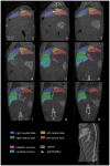
Figure 2. Coronally reconstructed micro-CT images.
Imaging of the liver of a C57BL/6J mouse was performed 3 hours after i.v. injection of 100 µl of a nanoparticular contrast agent (ExiTron nano 12000). The color-coding of the single liver lobes corresponds to the color coding presented in Figure 1. The slice orientation in relation to the spine is shown in the lower right corner.
Figure 3
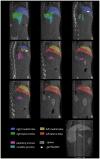
Figure 3. Saggital reconstructed micro-CT images.
Micro-CT of the liver of a C57BL/6J mouse was performed 3 hours after injection of 100 µl of a nanoparticular contrast agent (ExiTron nano 12000). The color-coding of the single liver lobes corrsponds to the color coding presented in Figure 1. The slice orientation in relation to a coronal section is shown in the lower right corner.
Figure 4
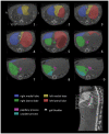
Figure 4. Axial reconstructed micro-CT images.
Micro-CT of the liver of a C57BL/6J mouse was performed 3 hours after injection of a nanoparticular contrast agent (ExiTron nano). The color-coding of the single liver lobes corrsponds to the color coding presented in Figure 1. The orientation of the axial slices in relation to the spine is shown in the lower right corner.
Figure 5
Figure 5. Liver variationes.
A–C are comparable coronal sections of the murine liver illustrating the variability of the size of the papillary process of the caudate lobe. D shows transdiaphragmatic herniation of parts of the right medial liver lobe, which we incidentally discovered during our studies.
Figure 6
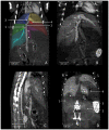
Figure 6. Perihepatic structures.
A shows coronally reconstructed micro-CT images acquired 20 minutes after injection of 400 µl Fenestra VC. The portal vein (arrowheads) runs in a caudocranial direction and divides into single veins supplying the liver lobes with blood from the abdominal organs. The right branch (1; Ramus dexter) divides up into two non-specified branches supplying the right lateral lobe (colored green) and the right caudate process (colored cyan). The left branch (2; Ramus sinister) divides into the umbillical part (3; Pars umbillicalis) and the transversal part (4; Pars transversalis) draining the left medial lobe and the left lateral lobe, respectively. The vein supplying the right medial liver lobe (colored blue) is not classified in the NAV. Spontaneous peristaltic movement of the portal vein is known to result in a corkscrew-like appearance of the vessel as shown in B. The liver is supplied with arterial blood from the coeliac artery (arrow) arising from the aorta cranial of the cranial mesenteric artery (arrowhead), as shown in C. The portal vein collecting blood from smaller abdominal vessels can be spotted directly ventral to the hepatic artery. D shows a coronally reconstructed micro-CT, 21 days after injection of 100 µl Exitron nano 12000 showing relevant organs adjacent to the liver. The right kidney (1), the left kidney (2), the spleen (3), the stomach (4; containing air bubbles), and the adrenal glands (arrow heads). The dotted lines indicate the renal impression of the liver on the right side and the gastric impression on the left side.
Figure 7
Figure 7. Hepatic veins.
Maximum intensity projection (MIP) of micro-CT images of the murine hepatic veins 20 minutes after injection of 400 µl Fenestra VC. The color coding of the draining veins corresponds to the color coding used for the liver lobes before. The renal veins are indicated by arrowheads.
Figure 8
Figure 8. Illustrative micro-CT images of liver metatstases in a mouse.
A–C Images of liver metastases were acquired 2 hours after i.v. injection of 100 µl of a contrast agent (Viscover Exitron nano 6000; Miltenyi Biotec, Bergisch-Gladbach, Germany) in a coronal (A), transversal (B), and saggital (C) slice orientation. With regard to liver anatomy, we found metastases to develop predominantly under the liver capsule adjacent to the fissures between the liver lobes. While the left lateral lobe (LLL) shows no metastases on its caudal edge in this slice, there are plenty on its cranial edge, next to the left medial lobe (LML). The lobes on the right side (right medial lobe (RML), right lateral lobe (RLL) and the caudate process (CP)) show (besides the subcapsular growth pattern) metastases within the liver parenchyma. Due to tumor growth the papillary process depicted in B and C lost its typical shape (compare with figs. 2, 3, 4).

Similar articles

Cited by

References

    1. Dorner M, Horwitz JA, Robbins JB, Barry WT, Feng Q, et al. A genetically humanized mouse model for hepatitis C virus infection. Nature. 2011;474:208–211. - PMC - PubMed
    1. Kuhara T, Yamauchi K, Iwatsuki K. Bovine lactoferrin induces interleukin-11 production in a hepatitis mouse model and human intestinal myofibroblasts. Eur J Nutr. 2011 in press. - PubMed
    1. Gabele E, Dostert K, Dorn C, Patsenker E, Stickel F, et al. A new model of interactive effects of alcohol and high-fat diet on hepatic fibrosis. Alcohol Clin Exp Res. 2011;35:1361–1367. - PubMed
    1. Yan AW, Fouts DE, Brandl J, Starkel P, Torralba M, et al. Enteric dysbiosis associated with a mouse model of alcoholic liver disease. Hepatology. 2011;53:96–105. - PMC - PubMed
    1. Van Steenkiste C, Staelens S, Deleye S, De Vos F, Vandenberghe S, et al. Measurement of porto-systemic shunting in mice by novel three-dimensional micro-single photon emission computed tomography imaging enabling longitudinal follow-up. Liver International. 2010;30:1211–1220. - PubMed

Publication types

MeSH terms