. 2012;7(2):e31188.
doi: 10.1371/journal.pone.0031188. Epub 2012 Feb 8.

Recombinant lysyl oxidase propeptide protein inhibits growth and promotes apoptosis of pre-existing murine breast cancer xenografts

Affiliations

Recombinant lysyl oxidase propeptide protein inhibits growth and promotes apoptosis of pre-existing murine breast cancer xenografts

Manish V Bais et al. PLoS One. 2012.

Abstract

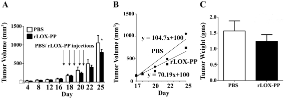
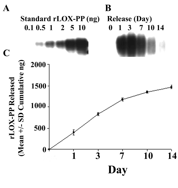
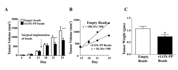
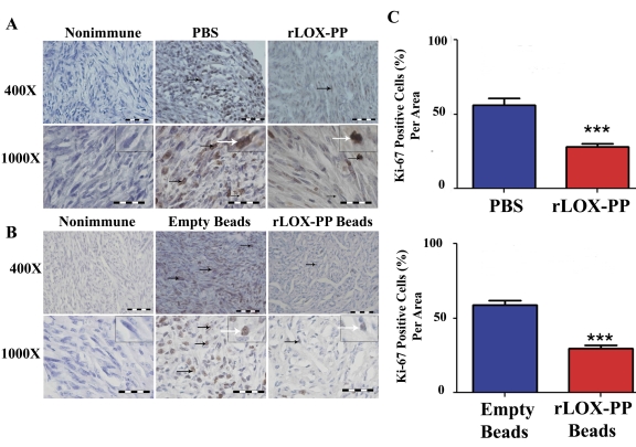
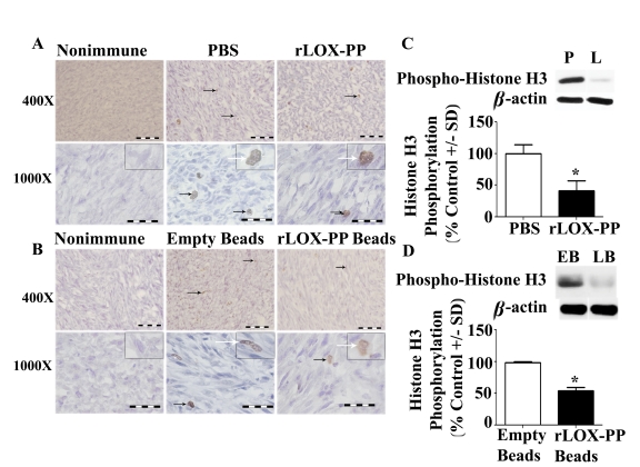
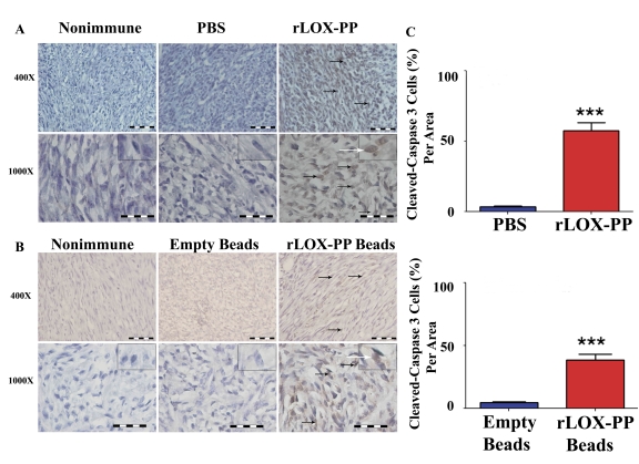
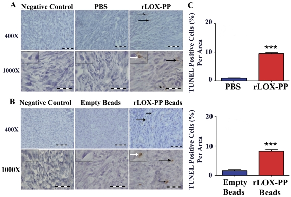
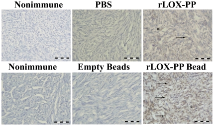
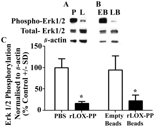
Lysyl oxidase propeptide (LOX-PP) ectopic overexpression inhibits the growth of cancer xenografts. Here the ability and mode of action of purified recombinant LOX-PP (rLOX-PP) protein to inhibit the growth of pre-existing xenografts was determined. Experimental approaches employed were direct intratumoral injection (i.t.) of rLOX-PP protein into murine breast cancer NF639 xenografts, and application of a slow release formulation of rLOX-PP implanted adjacent to tumors in NCR nu/nu mice (n = 10). Tumors were monitored for growth, and after sacrifice were subjected to immunohistochemical and Western blot analyses for several markers of proliferation, apoptosis, and for rLOX-PP itself. Direct i.t. injection of rLOX-PP significantly reduced tumor volume on days 20, 22 and 25 and tumor weight at harvest on day 25 by 30% compared to control. Implantation of beads preloaded with 35 micrograms rLOX-PP (n = 10) in vivo reduced tumor volume and weight at sacrifice when compared to empty beads (p<0.05). A 30% reduction of tumor volume on days 22 and 25 (p<0.05) and final tumor weight on day 25 (p<0.05) were observed with a reduced tumor growth rate of 60% after implantation. rLOX-PP significantly reduced the expression of proliferation markers and Erk1/2 MAP kinase activation, while prominent increases in apoptosis markers were observed. rLOX-PP was detected by immunohistochemistry in harvested rLOX-PP tumors, but not in controls. Data provide pre-clinical findings that support proof of principle for the therapeutic anti-cancer potential of rLOX-PP protein formulations.


Conflict of interest statement

Competing Interests: The authors have declared that no competing interests exist.

Figures

Figure 1
Figure 1. Injection of rLOX-PP inhibits NF639 xenograft growth in mice.
(A) Subcutaneous xenografts of NF639 breast cancer cells were injected once daily with 10 micrograms rLOX-PP for 5 days beginning on day 17 (arrows). Tumor volumes were monitored by caliper measurements (n = 10; *, p<0.05); (B) The tumor growth rates were determined from measurements made beginning on day 18 and were derived from linear regression analyses of the data. (C) Tumor weights at sacrifice on day 25 (p = 0.4, n = 10).
Figure 2
Figure 2. Alginate bead release kinetics of rLOX-PP.
(A) Western blot of known amounts of rLOX-PP ranging from 0.1 to 10 ng of rLOX-PP; (B) Western blot of rLOX-PP-bead supernatants collected at different intervals; (C) calculated cumulative release of rLOX-PP; (n = 4; *, p<0.001).
Figure 3
Figure 3. rLOX-PP incorporated in alginate beads inhibits NF639 xenograft growth in mice compared to empty beads.
(A) Subcutaneous 100 mm3 xenografts of NF639 breast cancer cells were established in mice. rLOX-PP/alginate or empty beads were implanted on day 15 (arrow). Tumors were monitored by caliper measurements (n = 10; *, p<0.05, ***, p<0.01). (B) The tumor growth rates were determined from measurements made beginning on day 18 and were derived from linear regression analyses of the data; (C) tumor weights determined at sacrifice on day 25 (n = 10; *, p<0.05).
Figure 4
Figure 4. Proliferation marker Ki-67 is decreased by rLOX-PP treatment.
(A) Immunostaining with Ki-67 antibody or non-immune IgG control tumors after (A) direct i.t. injection of rLOX-PP compared to PBS and (B) after implantation of empty and rLOX-PP alginate beads. Black arrows mark some positive-stained cells; the white arrows point to an inset containing an enlarged image of a stained cell. (C) The corresponding quantifications are shown (***, p<0.001; n = 3). Scale bar = 0.005 mm.
Figure 5
Figure 5. Proliferation marker phosphorylated histone H3 is decreased by rLOX-PP treatment.
(A) Immunostaining with phospho- histone H3 antibody or nonimmune IgG control tumors after (A) direct i.t. injection of rLOX-PP (L) compared to PBS (P) and (B) after implantation of empty (EB) and rLOX-PP (LB) alginate beads. Black arrows mark some positive-stained cells; the white arrows point to an inset containing an enlarged image of a stained cell. Scale bars correspond to 0.005 mm for 400× and 0.002 mm for 1000×. Arrows mark some positively stained cells. (C) Western blot analyses of 17 kDa phospho- histone H3, 36 kDa beta-actin and the corresponding quantifications of protein extracts from tumors after (C) direct i.t. injections of rLOX-PP compared to PBS and (D) after implantation of empty and rLOX-PP alginate beads (*, p<0.01; n = 3).
Figure 6
Figure 6. Apoptosis marker active caspase 3 is increased by rLOX-PP treatment.
(A) Immunostaining of tumors with active caspase 3 antibody, or non-immune IgG control. (A) i.t. rLOX-PP- or PBS injected tumors, and (B) tumors treated with surgically implanted empty alginate beads or rLOX-PP/alginate beads. Black arrows mark some positive-stained cells; the white arrows point to an inset containing an enlarged image of a stained cell. Scale bar = 0.005 mm for 400× and 0.002 mm for 1000×; (C) the corresponding quantifications (***, p<0.001; n = 3).
Figure 7
Figure 7. Apoptosis marker TUNEL is increased by rLOX-PP treatment.
(A) TUNEL staining of tumors treated i.t. with rLOX-PP, or tumors injected with negative control PBS; (B) tumors treated with surgically implanted empty alginate beads or rLOX-PP alginate beads. Black arrows mark some positive-stained cells; the white arrows point to an inset containing an enlarged image of a stained cell. Scale bar = 0.005 mm for 400× and 0.002 mm for 1000×; (C) the corresponding quantifications (***, p<0.001; n = 3). Arrows mark some positive-stained cells.
Figure 8
Figure 8. rLOX-PP persists in treated tumors.
Immunostaining of tumors after sacrifice with LOX-PP antibody or non-immune IgG control from mice after (A) direct i.t. injection of rLOX-PP compared to PBS or (B) after implantation of empty or rLOX-PP-containing alginate beads. Scale bars = 0.005 mm. Arrows mark some positive-stained cells.
Figure 9
Figure 9. LOX-PP inhibits Ras effector Erk1/2 phosphorylation in vivo.
Western blot analysis of 42 and 44 kDa phospho-Erk, total Erk1/2 and 36 kDa beta-actin from tumor extracts after (A) direct i.t. injections of rLOX-PP (L) compared to PBS (P); (B) after implantation of empty (EB) and rLOX-PP (LB) alginate beads; (C) the corresponding quantifications are shown (*, p<0.05; n = 3).

Similar articles

Cited by

References

    1. Kagan HM, Trackman PC. Properties and function of lysyl oxidase. Am J Respir Cell Mol Biol. 1991;5:206–210. - PubMed
    1. Cronshaw AD, Fothergill-Gilmore LA, Hulmes DJ. The proteolytic processing site of the precursor of lysyl oxidase. Biochem J. 1995;306(Pt 1):279–284. - PMC - PubMed
    1. Kessler E, Takahara K, Biniaminov L, Brusel M, Greenspan DS. Bone morphogenetic protein-1: the type I procollagen C-proteinase. Science. 1996;271:360–362. - PubMed
    1. Uzel MI, Scott IC, Babakhanlou-Chase H, Palamakumbura AH, Pappano WN, et al. Multiple bone morphogenetic protein 1-related mammalian metalloproteinases process pro-lysyl oxidase at the correct physiological site and control lysyl oxidase activation in mouse embryo fibroblast cultures. J Biol Chem. 2001;276:22537–22543. - PubMed
    1. Palamakumbura AH, Jeay S, Guo Y, Pischon N, Sommer P, et al. The propeptide domain of lysyl oxidase induces phenotypic reversion of ras-transformed cells. J Biol Chem. 2004;279:40593–40600. - PubMed

Publication types

MeSH terms