. 2012;7(2):e31255.
doi: 10.1371/journal.pone.0031255. Epub 2012 Feb 17.

Activation of the PI3K/AKT pathway in Merkel cell carcinoma

Affiliations

Activation of the PI3K/AKT pathway in Merkel cell carcinoma

Christian Hafner et al. PLoS One. 2012.

Abstract

Merkel cell carcinoma (MCC) is a highly aggressive skin cancer with an increasing incidence. The understanding of the molecular carcinogenesis of MCC is limited. Here, we scrutinized the PI3K/AKT pathway, one of the major pathways activated in human cancer, in MCC. Immunohistochemical analysis of 41 tumor tissues and 9 MCC cell lines revealed high levels of AKT phosphorylation at threonine 308 in 88% of samples. Notably, the AKT phosphorylation was not correlated with the presence or absence of the Merkel cell polyoma virus (MCV). Accordingly, knock-down of the large and small T antigen by shRNA in MCV positive MCC cells did not affect phosphorylation of AKT. We also analyzed 46 MCC samples for activating PIK3CA and AKT1 mutations. Oncogenic PIK3CA mutations were found in 2/46 (4%) MCCs whereas mutations in exon 4 of AKT1 were absent. MCC cell lines demonstrated a high sensitivity towards the PI3K inhibitor LY-294002. This finding together with our observation that the PI3K/AKT pathway is activated in the majority of human MCCs identifies PI3K/AKT as a potential new therapeutic target for MCC patients.


Conflict of interest statement

Competing Interests: The authors have declared that no competing interests exist.

Figures

Figure 1
Figure 1. Activating phosphorylation of the AKT protein at position threonine 308 in Merkel cell carcinoma, malignant melanoma and basal cell carcinoma.
The presence of AKT phosphorylated at T308 was analyzed by immunohistochemistry using a phospho-specific antibody on tissue micro arrays representing 41 MCCs (each in triplicates), 45 basal cell carcinomas (BCC) and 67 melanomas, respectively. Samples were scored from 0 (negative) to 3+ (strongly positive). The percentage of the samples for each expression score is indicated as bar graph and examples for the staining intensity in MCC are depicted below. In total, 88% of MCC samples showed strong (+2) or very strong (+3) staining for phospho-AKT T308, while 59% of melanoma samples were observed in these categories. Basal cell carcinoma showed only negative or weak AKT phosphorylation.
Figure 2
Figure 2. Merkel cell polyomavirus T antigens do not affect AKT phosphorylation in Merkel cell carcinoma.
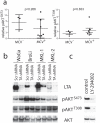
a) Total cell lysates of 7 MCV positive and 3 MCV negative MCC cell lines were subjected to Western blot analysis applying antibodies to pAKTT308, pAKTS473 and tubulin. Signal intensity was quantified using the imageJ software and the values normalized p-values according to the Mann-Whitney are indicated. b) The indicated cell lines were infected with the lentiviral shRNA vector KH1 encoding GFP and either a shRNA targeting all MCV TA mRNAs or a scrambled shRNA; Infection rates as determined by GFP flow cytometry analysis were 98% for WaGa, 94% for BroLi, 90% for MKL-1 and 96% for MKL-2. Total cell lysates harvested on day 5 following infection were then analyzed by immunoblotting for expression of large T antigen (LTA) and AKT and for the presence of AKT phosphorylated at T308 or S473. The variations in molecular size of the LTA proteins in the different cell lines are due to different stop codon mutations truncating the C-terminal part of the protein. c) Treatment with the PI-3 kinase inhibitor LY-294002 demonstrated inhibition of AKT phosphorylation at the phosphorylation sites T308 and S473 by blocking the upstream kinase.
Figure 3
Figure 3. PIK3CA hotspot mutations in Merkel cell carcinoma.
(a) The heterozygous p.E542K (c.G1624A) PIK3CA hotspot mutation was detected in sample no. 27 by direct sequencing (left) and a PIK3CA SNaPshot® assay (right) covering the most frequent hotspot PIK3CA mutations. The number of the wildtype codons is indicated above the peaks in the SNaPshot® assay. In brief, the SNaPshot® assay comprises a multiplex PCR for exons 9 and 20 of PIK3CA, followed by extension of 4 primers specific for the most frequent PIK3CA hotspot mutation loci. Because fluorescent dideoxynucleotides are used for this primer extension step, only one peak appears at the base position with the potential mutation. The color of the peak allows discrimination of wildtype and mutated alleles. (b) The heterozygous p.E545Q (c.G1633C) PIK3CA hotspot mutation was detected in sample no. 24 by direct sequencing (left) and a PIK3CA SNaPshot® assay (right) covering the most frequent hotspot PIK3CA mutations. The number of the respective wildtype codon is indicated above the peaks in the SNaPshot® assay.
Figure 4
Figure 4. MCC cell lines are sensitive to the PI3K inhibitor LY-294002.
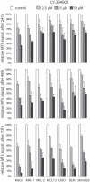
The indicated MCC and melanoma cell lines were incubated with LY-294002 at three different concentrations. After the indicated time period the cells were subjected to the MTS assay in triplicates. The reduction in extinction relative to the DMSO (solvent of LY-294002) controls is depicted. The graphs represent mean values (± standard deviation) of at least three independent experiments.

Similar articles

Cited by

References

    1. Becker JC, Schrama D, Houben R. Merkel cell carcinoma. Cell Mol Life Sci. 2009;66:1–8. - PMC - PubMed
    1. Becker JC, Houben R, Ugurel S, Trefzer U, Pfohler C, et al. MC polyomavirus is frequently present in Merkel cell carcinoma of European patients. J Invest Dermatol. 2009;129:248–250. - PubMed
    1. Feng H, Shuda M, Chang Y, Moore PS. Clonal integration of a polyomavirus in human Merkel cell carcinoma. Science. 2008;319:1096–1100. - PMC - PubMed
    1. Houben R, Shuda M, Weinkam R, Schrama D, Feng H, et al. Merkel cell polyomavirus infected Merkel cell carcinoma cells require expression of viral T antigens. J Virol. 2010;84:7064–7072. - PMC - PubMed
    1. Lemos B, Nghiem P. Merkel cell carcinoma: more deaths but still no pathway to blame. J Invest Dermatol. 2007;127:2100–2103. - PubMed

Publication types

MeSH terms

Substances