. 2012;7(2):e31296.
doi: 10.1371/journal.pone.0031296. Epub 2012 Feb 21.

Preformed CD40L is stored in Th1, Th2, Th17, and T follicular helper cells as well as CD4+ 8- thymocytes and invariant NKT cells but not in Treg cells

Affiliations

Preformed CD40L is stored in Th1, Th2, Th17, and T follicular helper cells as well as CD4+ 8- thymocytes and invariant NKT cells but not in Treg cells

Yoshinobu Koguchi et al. PLoS One. 2012.

Abstract

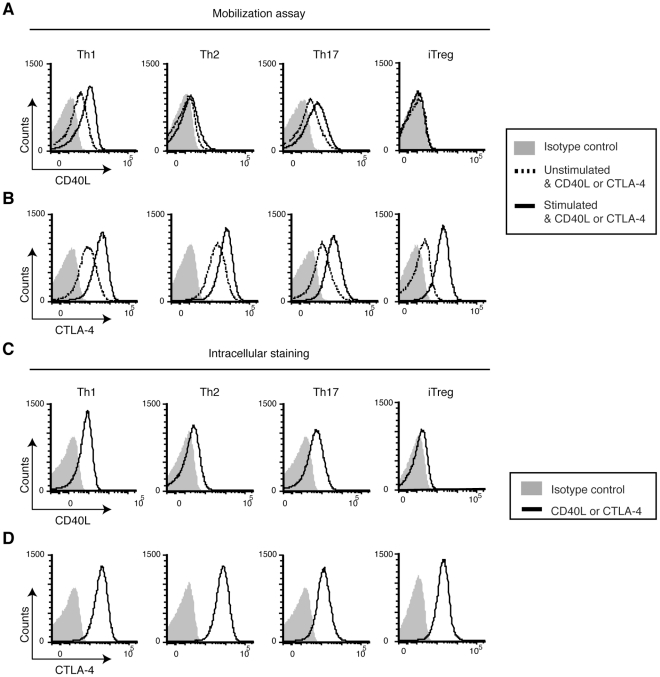
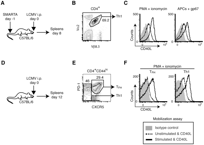
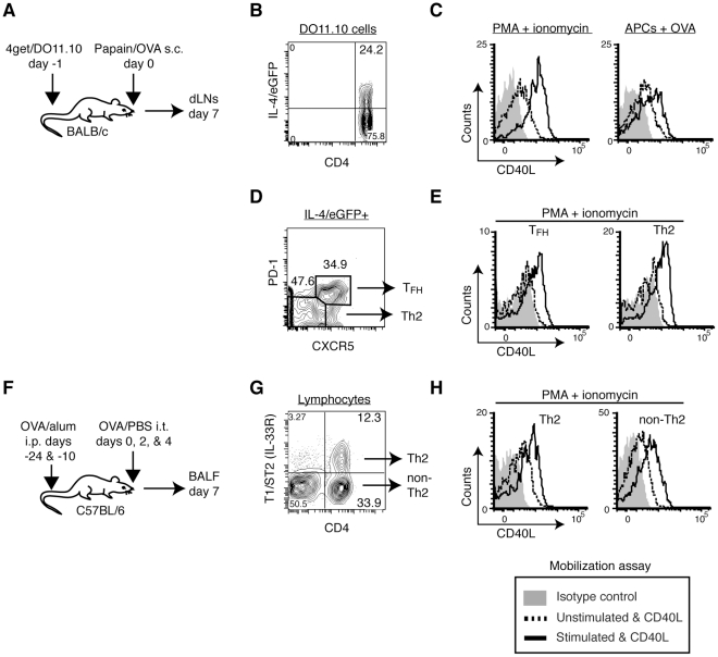
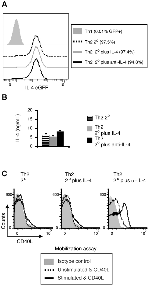
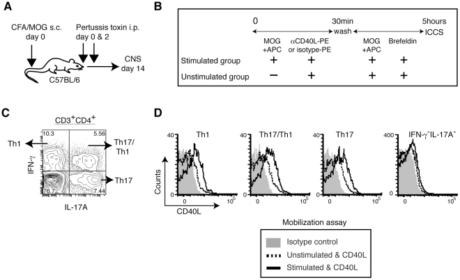
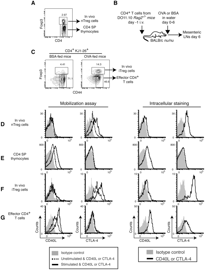
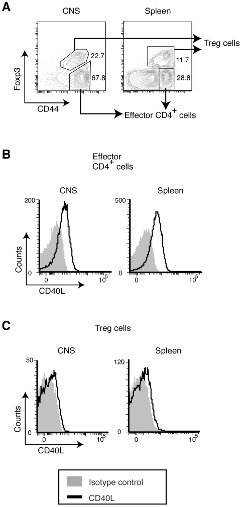
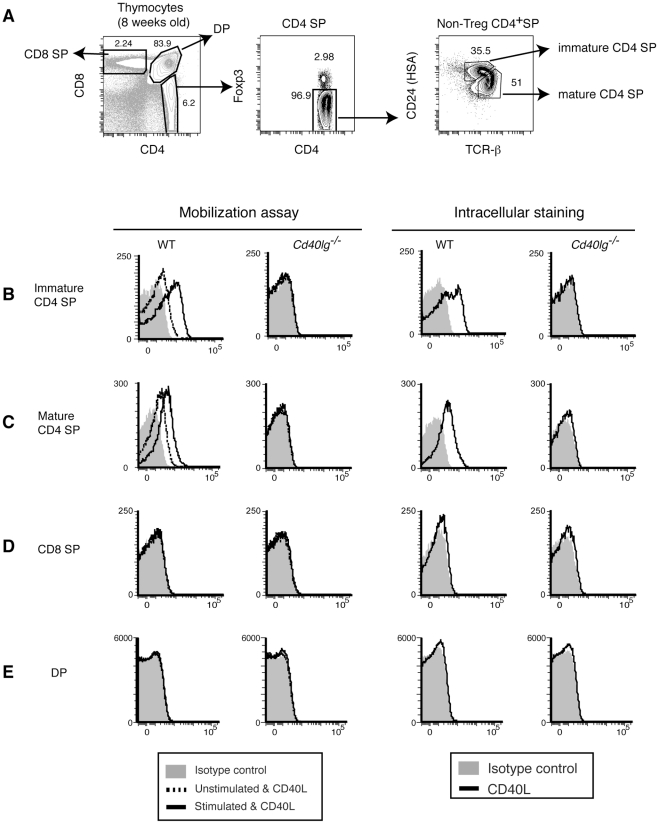
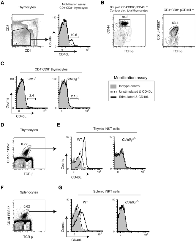
CD40L is essential for the development of adaptive immune responses. It is generally thought that CD40L expression in CD4(+) T cells is regulated transcriptionally and made from new mRNA following antigen recognition. However, imaging studies show that the majority of cognate interactions between effector CD4(+) T cells and APCs in vivo are too short to allow de novo CD40L synthesis. We previously showed that Th1 effector and memory cells store preformed CD40L (pCD40L) in lysosomal compartments and mobilize it onto the plasma membrane immediately after antigenic stimulation, suggesting that primed CD4(+) T cells may use pCD40L to activate APCs during brief encounters. Indeed, our recent study showed that pCD40L is sufficient to mediate selective activation of cognate B cells and trigger DC activation in vitro. In this study, we show that pCD40L is present in Th1 and follicular helper T cells developed during infection with lymphocytic choriomeningitis virus, Th2 cells in the airway of asthmatic mice, and Th17 cells from the CNS of animals with experimental autoimmune encephalitis (EAE). pCD40L is nearly absent in both natural and induced Treg cells, even in the presence of intense inflammation such as occurs in EAE. We also found pCD40L expression in CD4 single positive thymocytes and invariant NKT cells. Together, these results suggest that pCD40L may function in T cell development as well as an unexpectedly broad spectrum of innate and adaptive immune responses, while its expression in Treg cells is repressed to avoid compromising their suppressive activity.


Conflict of interest statement

Competing Interests: The authors have declared that no competing interests exist.

Figures

Figure 1
Figure 1. In vitro-generated Th1 and Th17, but not Th2 or iTreg cells mobilize pCD40L.
A and B, Mobilization assay. In vitro-generated Th1, Th2, Th17, and iTreg cells were stimulated with PMA plus ionomycin or left unstimulated in the presence of PE-isotype Ab, PE-anti-CD40L or PE-anti-CTLA-4 at 37°C for 30 minutes. The levels of CD40L (A) and CTLA-4 (B) are shown. C and D, Intracellular staining. Cells were fixed without stimulation, permeabilized, and stained with PE-isotype Ab, PE-anti-CD40L or PE-anti-CTLA-4. The levels of CD40L (C) and CTLA-4 (D) are shown. Data are representative of five independent experiments.
Figure 2
Figure 2. In vivo-generated Th1 and TFH cells possess and mobilize pCD40L.
A, Generation of antigen-specific Th1 cells. LCMV-glycoprotein-specific, TCR-transgenic SMARTA CD4+ T cells were recovered from LCMV-infected mice on day 8 post-infection. B, Gating strategy for SMARTA cells (Vα2+, Vβ8.3+). C, Mobilization of pCD40L by SMARTA cells in response to PMA plus ionomycin or the LCMV peptide gp67-pulsed APC. D, Polyclonal Th1 and TFH cells. Spleen cells were obtained from LCMV-infected mice on day 12 post-infection. E, Gating strategy for TFH (CD4+CD44hiCXCR5hiPD-1hi) and Th1 (CD4+CD44hiCXCR5lowPD-1low) cells. F, Mobilization of pCD40L by TFH and Th1 cells in response to PMA plus ionomycin. Data are representative of two independent experiments.
Figure 3
Figure 3. In vivo-generated Th2 cells store and mobilize pCD40L.
A, BALB/c mice that had received 4get/DO11.10 CD4+ T cells were subcutaneously immunized with papain plus OVA protein. Seven days later, cells from dLNs were analyzed by the mobilization assay. B, Gating strategy for antigen-specific IL-4/eGFP+ DO11.10 cells (CD4+IL-4/eGFP+KJ1-26+). C, Mobilization of pCD40L by IL-4/eGFP+ DO11.10 cells in response to PMA plus ionomycin or OVA peptide-pulsed APC. D, Those cells were further differentiated as TFH (CXCR5hiPD-1hi) and Th2 (CXCR5lowPD-1low) cells. E, Mobilization of pCD40L by TFH and Th2 cells in response to PMA plus ionomycin. F, To obtain polyclonal Th2 cells, mice were sensitized with OVA/alum followed by intratracheal challenge with OVA/PBS. G, The gating strategy for Th2 cells (CD4+T1/ST2+) and non-Th2 cells in BALF. H, Mobilization of pCD40L by polyclonal Th2 and non-Th2 cells in response to PMA plus ionomycin. Data are representative of two to three independent experiments.
Figure 4
Figure 4. Excess IL-4 downregulates pCD40L levels in Th2 cells generated in vitro.
Recovery of pCD40L in Th2 cells by neutralizing IL-4. A–C, Differentiated Th2 cells from 4get/DO11.10 spleen cells were restimulated with peptide-pulsed splenic APCs alone (Th2 2°) or in the presence of exogenous IL-4 (Th2 2° plus IL-4) or anti-IL-4 (Th2 2° plus anti-IL-4) for 4 days. A, The levels of IL-4/eGFP in each group of Th2 cells as well as Th1 negative control cells. B, IL-4 production upon PMA plus ionomycin stimulation by each group of Th2 cells was analyzed by ELISA. Each bar represents the mean ± standard deviation for triplicates. C, Mobilization of pCD40L by each group of Th2 cells in response to PMA plus ionomycin. Data are representative of two independent experiments.
Figure 5
Figure 5. In vivo-generated Th17 and Th17/Th1 cells store and mobilize pCD40L.
A, CNS infiltrating leukocytes were obtained from brains and spinal cords of EAE animals induced by immunization with CFA plus MOG peptide and pertussis toxin. B, CNS infiltrating leukocytes were analyzed by the CD40L mobilization assay followed by intracellular cytokine staining. C, Intracellular staining of IL-17A and IFN-γ in CNS CD4+ T cells. D, CD40L mobilization in Th1 (IL-17AIFN-γ+), Th17 (IL-17A+IFN-γ), Th17/Th1 (IL-17A+IFN-γ+), and antigen non-specific (IL-17AIFN-γ) CD4+ T cells upon antigenic stimulation. Data are representative of three independent experiments.
Figure 6
Figure 6. CD4 SP thymocytes, but not Treg cells, store and mobilize pCD40L.
A, Gating strategy for thymic nTreg cells and CD4 SP thymocytes. B, Generation of in vivo iTreg cells. DO11.10 CD4+ T cells were recovered from OVA- or BSA-fed recipient mice on day 6. C, Gating strategy for in vivo iTreg cells and effector CD4+ T cells. A BSA-fed mouse is shown as a negative control. D, thymic nTreg cells; E, CD4 SP thymocytes; F, in vivo iTreg cells; and G, effector CD4+ T cells, are analyzed by the mobilization assay and intracellular staining for pCD40L and CTLA-4. Data are representative of three (D and E) or two (F and G) independent experiments.
Figure 7
Figure 7. Severe inflammation does not compromise the lack of expression of pCD40L by Treg cells.
CNS infiltrating leukocytes and splenocytes were obtained from EAE animals induced as Fig. 5A . A, Gating strategy for cells from CNS and spleen. B & C, Intracellular CD40L levels for effector CD4+ T cells (B) and Treg cells (C). Data are representative of two independent experiments.
Figure 8
Figure 8. CD4 SP thymocytes, but not CD8 SP or DP thymocytes, store and mobilize pCD40L.
A, Gating strategy for identifying CD8 SP, DP, and immature and mature CD4 SP thymocytes. B–E, Mobilization of pCD40L for immature (B) and mature (C) CD4 SP thymocytes, CD8 SP (D), and DP (E) thymocytes. Cd40lg−/− : CD40L-deficient mouse. Data are representative of at least five independent experiments.
Figure 9
Figure 9. iNKT cells possess and mobilize pCD40L.
A, A fraction of the DN thymocyte population mobilizes pCD40L upon stimulation with PMA plus ionomycin. B, pCD40L positive DN thymocytes are iNKT cells. C, iNKT deficient β2m−/− mice lack the pCD40L positive DN thymocyte population. D and F, Gating strategy for thymic (D) and splenic (F) iNKT cells. E and G, Mobilization of pCD40L in CD1d-tetramer-positive thymic (E) or splenic (G) iNKT cells upon stimulation with PMA plus ionomycin. Data are representative of two independent experiments.

Similar articles

Cited by

References

    1. McHeyzer-Williams LJ, McHeyzer-Williams MG. Antigen-specific memory B cell development. Annu Rev Immunol. 2005;23:487–513. - PubMed
    1. Williams MA, Bevan MJ. Effector and memory CTL differentiation. Annu Rev Immunol. 2007;25:171–192. - PubMed
    1. Parker DC. T cell-dependent B cell activation. Annu Rev Immunol. 1993;11:331–360. - PubMed
    1. Feau S, Arens R, Togher S, Schoenberger SP. Autocrine IL-2 is required for secondary population expansion of CD8(+) memory T cells. Nat Immunol. 2011;12:908–913. - PMC - PubMed
    1. van Kooten C, Banchereau J. CD40-CD40 ligand. J Leukoc Biol. 2000;67:2–17. - PubMed

Publication types

MeSH terms