. 2012;7(2):e31336.
doi: 10.1371/journal.pone.0031336. Epub 2012 Feb 17.

Neonatal periostin knockout mice are protected from hyperoxia-induced alveolar simplication

Affiliations

Neonatal periostin knockout mice are protected from hyperoxia-induced alveolar simplication

Paul D Bozyk et al. PLoS One. 2012.

Erratum in

Abstract

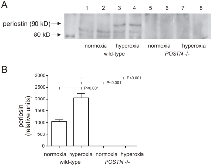
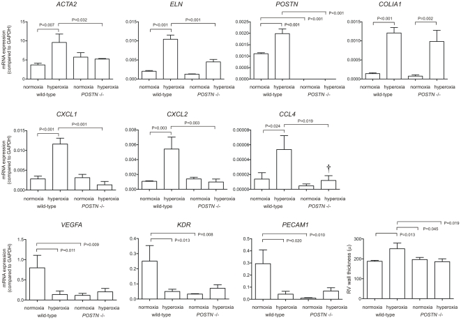
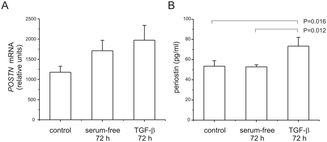
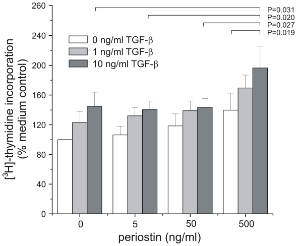
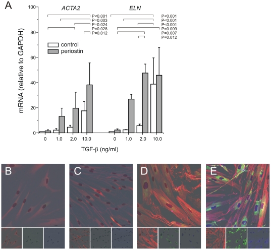
In bronchopulmonary dysplasia (BPD), alveolar septae are thickened with collagen and α-smooth muscle actin, transforming growth factor (TGF)-β-positive myofibroblasts. Periostin, a secreted extracellular matrix protein, is involved in TGF-β-mediated fibrosis and myofibroblast differentiation. We hypothesized that periostin expression is required for hypoalveolarization and interstitial fibrosis in hyperoxia-exposed neonatal mice, an animal model for this disease. We also examined periostin expression in neonatal lung mesenchymal stromal cells and lung tissue of hyperoxia-exposed neonatal mice and human infants with BPD. Two-to-three day-old wild-type and periostin null mice were exposed to air or 75% oxygen for 14 days. Mesenchymal stromal cells were isolated from tracheal aspirates of premature infants. Hyperoxic exposure of neonatal mice increased alveolar wall periostin expression, particularly in areas of interstitial thickening. Periostin co-localized with α-smooth muscle actin, suggesting synthesis by myofibroblasts. A similar pattern was found in lung sections of infants dying of BPD. Unlike wild-type mice, hyperoxia-exposed periostin null mice did not show larger air spaces or α-smooth muscle-positive myofibroblasts. Compared to hyperoxia-exposed wild-type mice, hyperoxia-exposed periostin null mice also showed reduced lung mRNA expression of α-smooth muscle actin, elastin, CXCL1, CXCL2 and CCL4. TGF-β treatment increased mesenchymal stromal cell periostin expression, and periostin treatment increased TGF-β-mediated DNA synthesis and myofibroblast differentiation. We conclude that periostin expression is increased in the lungs of hyperoxia-exposed neonatal mice and infants with BPD, and is required for hyperoxia-induced hypoalveolarization and interstitial fibrosis.


Conflict of interest statement

Competing Interests: The authors have declared that no competing interests exist.

Figures

Figure 1
Figure 1. Hyperoxia induces a BPD phenotype in neonatal mice.
Two-to-three day-old wild-type C57BL/6J mice were exposed to air or 75% oxygen for 14 days. Compared to air-exposed mice (panels A–D). hyperoxic exposure caused the development of fewer and larger airspaces (E). Fluorescence microscopy showed increased deposition of α-actin (red), elastin (green) and collagen-I (blue, F). Colocalization of α-actin, elastin and collagen-I appears white (arrow, inset). G. Immunohistochemical stains showed periostin expression in the alveolar walls, particularly in areas of interstitial thickening. H. Periostin expression (green) colocalized with α-smooth muscle actin (red) and collagen (blue), suggesting synthesis by myofibroblasts Colocalization of periostin and collagen appears light blue; colocalization of α-actin, periostin and collagen-I appears white (arrow, inset). Colocalization was also present at the tips of secondary crests (arrowhead). Original magnification, 200×. These results are typical of three individual experiments.
Figure 2
Figure 2. Hyperoxic exposure on neonatal mice increases lung periostin content.
Whole lung lysates were resolved by SDS-PAGE, transferred to nitrocellulose and probed with anti-periostin (A). Results from two normoxic wild-type mice and two hyperoxic wild-type mice are shown here; lysates from normoxic and hyperoxic periostin null mice are added for comparison. Immunoblots showed a full-length 90 kD isoform as well as two smaller bands. (B). Group mean data showing a significant increase in periostin expression with hyperoxic exposure (n = 4 for each group, one way ANOVA).
Figure 3
Figure 3. Increased periostin expression in the lungs of infants with BPD.
The lung of a full-term infants dying of a non-pulmonary cause is shown in (A). There is significant staining in the airway subepithelium, with miminal staining of the airway epithelium or alveolar walls. B–D. Staining of lung sections from three individual infants dying of BPD showed increased periostin expression, particularly in the subepithelium and fibroblastic foci. E. We also examined periostin (green) and α-smooth muscle actin (red) expression by fluorescence microscopy. Lungs of full-term infants showed periostin expression in the airway subepithelium which was distinct from the adjacent smooth muscle. F–H. Lungs of three individual infants with BPD were also examined for periostin expression. Lungs showed colocalization of periostin and α-actin in interstitial alveolar myofibroblasts (F and G, arrows, insets). Colocalization of periostin and α-actin (yellow-orange) was also found at the tips of secondary crests (H, arrow, inset). Original magnification, 200×.
Figure 4
Figure 4. Periostin knockout prevents hypoventilation and myofibroblast differentiation in hyperoxia-exposed neonatal mice.
Two-day-old periostin null mice were exposed to air or 75% oxygen for 14 days. Compared to air-exposed wild-type mice (A), hyperoxia-exposed wild-type mice showed alveolar simplication (panel B). In contrast, air- (C) and hyperoxia-exposed periostin null mice (D) showed normal alveolar architecture. (E). Hypoalveolarization in wild-type hyperoxia-exposed mice was associated with a statistically significant increase in mean alveolar chord length (n = 4, one-way ANOVA).
Figure 5
Figure 5. Hyperoxic exposure is associated with α-actin and periostin-double positive myofibroblasts in wild-type but not periostin null mice.
Lung sections were stained for α-actin (red), periostin (green) and collagen I (blue); colocalization appears white. Unlike air-exposed wild-type mice (panel A), hyperoxia-exposed wild-type mice showed thickening of the interstitial space with α-smooth muscle-, periostin- and collagen type I-positive myofibroblasts (B). Air- (C) and hyperoxia-exposed periostin null mice (D) did not show alveolar myofibroblasts. These results are typical of three individual experiments.
Figure 6
Figure 6. Effects of hyperoxia on lung mRNA expression in wild-type and periostin null mice.
mRNA was measured by quantitative PCR. We examined mRNA expression of ACTA2, ELN, POSTN, COLIA1, CXCL1, CXCL2, CCL4, VEGFA, KDR and PECAM1 (n = 4, one-way ANOVA). Mice were also analyzed by right ventricular wall thickness (n = 4–6, one way ANOVA).
Figure 7
Figure 7. TGF-β treatment of mesenchymal stromal cells induces periostin expression.
Neonatal lung mesenchymal stromal cells were treated with 10 ng/ml TGF-β for 72 h and periostin mRNA and protein expression assessed by qPCR and ELISA, respectively. A. mRNA expression tended to increase with TGF-β treatment (n = 4). B. TGF-β treatment significantly increased periostin protein abundance (n = 7, one-way ANOVA).
Figure 8
Figure 8. Effects of periostin and TGF-β on mesenchymal stromal cell DNA synthesis.
Mesenchymal stromal cells (n = 4) were incubated with [3H]-thymidine and treated with either TGF-β or periostin. [3H]-thymidine incorporation was assessed by scintillation counting. We used two-way ANOVA with Fisher's least significant difference multiple comparison test to assess the individual and combined effects of TGF-β and periostin on neonatal lung mesenchymal stromal cell DNA synthesis. In the presence of 500 ng/ml periostin, 10 ng/ml TGF-β significantly increased DNA synthesis compared to periostin alone. Also, in the presence of 10 ng/ml TGF-β, 500 ng/ml periostin significantly increased DNA synthesis compared to other periostin concentrations.
Figure 9
Figure 9. Effects of periostin and TGF-β on mesenchymal stromal cell myofibroblastic differentiation.
A. Periostin (50 ng/ml) was incubated with mesenchymal stromal cells (n = 6) in the presence or absence of TGF-β (2–10 ng/ml). α-smooth muscle actin and elastin mRNA expression were measured by qPCR. We used two-way ANOVA with Fisher's least significant difference multiple comparison test to assess the individual and combined effects of TGF-β and periostin on neonatal lung mesenchymal stromal cell α-actin and elastin gene expression. TGF-β significantly increased α-actin and elastin expression in both the presence and absence of periostin. However, periostin significantly increased α-actin and elastin expression only in the presence of TGF-β. In other experiments, cells were stained with anti-α-smooth muscle actin (red), anti-collagen I (green) and anti-elastin (blue) (B, no treatment; C, periostin, 50 ng/ml; D, TGF-β, 2 ng/ml; E, periostin, 50 ng/ml and TGF-β, 2 ng/ml). Merged and individual images are shown (original magnification, 200×, results are representative of three individual experiments).

References

    1. Jobe AH, Bancalari E. Bronchopulmonary dysplasia. Am J Respir Crit Care Med. 2001;163:1723–1729. - PubMed
    1. Hussain NA, Siddiqui NH, Stocker JR. Pathology of arrested acinar development in postsurfactant bronchopulmonary dysplasia. Hum Pathol. 1998;29:710–717. - PubMed
    1. Coalson JJ. Pathology of new bronchopulmonary dysplasia. Semin Neonatol. 2003;8:73–81. - PubMed
    1. Toti P, Buonocore G, Tanganelli P, Catella AM, Palmeri ML, et al. Bronchopulmonary dysplasia of the premature baby: an immunohistochemical study. Pediatr Pulmonol. 1997;24:22–28. - PubMed
    1. Bhatt AJ, Pryhuber GS, Huyck H, Watkins RH, Metlay LA, et al. Disrupted pulmonary vasculature and decreased vascular endothelial growth factor, Flt-1, and TIE-2 in human infants dying with bronchopulmonary dysplasia. Am J Respir Crit Care Med. 2001;164:1971–1980. - PubMed

Publication types

MeSH terms

LinkOut - more resources