. 2012;7(2):e31692.
doi: 10.1371/journal.pone.0031692. Epub 2012 Feb 20.

Transcriptional repressor NIR functions in the ribosome RNA processing of both 40S and 60S subunits

Affiliations

Transcriptional repressor NIR functions in the ribosome RNA processing of both 40S and 60S subunits

Jianguo Wu et al. PLoS One. 2012.

Abstract

Background: NIR was identified as an inhibitor of histone acetyltransferase and it represses transcriptional activation of p53. NIR is predominantly localized in the nucleolus and known as Noc2p, which is involved in the maturation of the 60S ribosomal subunit. However, how NIR functions in the nucleolus remains undetermined. In the nucleolus, a 47S ribosomal RNA precursor (pre-rRNA) is transcribed and processed to produce 18S, 5.8S and 28S rRNAs. The 18S rRNA is incorporated into the 40S ribosomal subunit, whereas the 28S and 5.8S rRNAs are incorporated into the 60S subunit. U3 small nucleolar RNA (snoRNA) directs 18S rRNA processing and U8 snoRNA mediates processing of 28S and 5.8 S rRNAs. Functional disruption of nucleolus often causes p53 activation to inhibit cell proliferation.

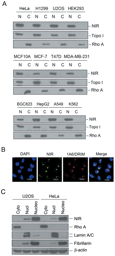
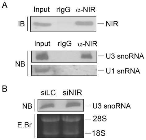
Methodology/principal findings: Western blotting showed that NIR is ubiquitously expressed in different human cell lines. Knock-down of NIR by siRNA led to inhibition of the 18S, 28S and 5.8S rRNAs evaluated by pulse-chase experiment. Pre-rRNA particles (pre-rRNPs) were fractionated from the nucleus by sucrose gradient centrifugation and analysis of the pre-RNPs components showed that NIR existed in the pre-RNPs of both the 60S and 40S subunits and co-fractionated with 32S and 12S pre-rRNAs in the 60S pre-rRNP. Protein-RNA binding experiments demonstrated that NIR is associated with the 32S pre-rRNA and U8 snoRNA. In addition, NIR bound U3 snoRNA. It is a novel finding that depletion of NIR did not affect p53 protein level but de-repressed acetylation of p53 and activated p21.

Conclusions: We provide the first evidence for a transcriptional repressor to function in the rRNA biogenesis of both the 40S and 60S subunits. Our findings also suggested that a nucleolar protein may alternatively signal to p53 by affecting the p53 modification rather than affecting p53 protein level.


Conflict of interest statement

Competing Interests: The authors have declared that no competing interests exist.

Figures

Figure 1
Figure 1. NIR is expressed in different human cell lines and mainly localized to the nucleolus.
A. Cytosolic and nuclear extracts were fractionized from multiple cancer cell lines as indicated at the top of blot. Same amount of protein was separated on SDS-PAGE, and transferred onto PVDF membranes. Blots were probed with anti-NIR. Fractionation was controlled by using nuclear marker protein topoisomerase I (Topo I) and a cytosolic marker protein RhoA. N represents nuclear extract and C represents cytoplasmic extract. B. Indirect immunofluorescence was performed with anti-NIR polyclonal antibody. NIR specific signal was recognized with FITC-conjugated goat anti-rabbit IgG. As a nucleolar protein, 1A6/DRIM was detected with anti-1A6/DRIM monoclonal antibody. 1A6/DRIM specific signal was recognized with TRITC-conjugated goat anti-mouse IgG. Nucleus was stained with DAPI. The image was obtained with confocal microscopy. C. Cytoplasmic, nucleoplasmic and nucleolar lysates were fractionized from U2OS and HeLa cells respectively. Same amount of protein from the above lysates was separated on SDS-PAGE, and transferred onto PVDF membranes. Blots were probed with anti-NIR. Rho A, lamin A/C and fibrillarin were used as controls for cytoplasmic, nucleoplasmic and nucleolar fractions respectively. Beta-actin was used as a loading control.
Figure 2
Figure 2. Knockdown of NIR resulted in inhibition of 18S, 28S and 5.8S rRNA processing.
A. Schematic representation of rRNA processing pathways in HeLa cells. B. U2OS cells were transfected with a NIR-specific siRNA (siNIR-1) or luciferase siRNA (siLC) as a control. At 72 hrs post-transfection, cell lysates were prepared. Proteins from the lysates were separated on SDS-PAGE, transferred onto a PVDF membrane. Blot was probed for detection of NIR protein (upper panel). NS, non-specific band. Above siRNA transfected U2OS cells were labeled with L-[methyl-3H] methionine and RNA was extracted at indicated time points. Equal amount of RNA from each sample was resolved on a 1% agarose gel. The gel was stained with ethidium bromide (EB) for photography (lower left panel) and transferred to a membrane for autoradiography (lower right panel). C. U2OS cells were transfected with siRNAs as in B. At 72 hrs post-transfection, cells were labeled with 3 µCi/ml [5, 6-3H] uridine for 30 min. RNA was extracted at indicated time points. Equal amount of RNA from each sample was resolved on a 10% polyacrylamide/7.5 M urea gel. The gel was stained with ethidium bromide (EB) for photography (left panel) and transferred to a membrane for autoradiography (right panel). D. U2OS cells were transfected with a second NIR-specific siRNA (siNIR-2) or luciferase siRNA (siLC) as a control. At 72 hrs post-transfection, cell lysates were prepared and subjected to Western blotting for detection of NIR protein (upper panel). Beta-actin was used as a loading control. Above siRNAs (siNIR-2 and siLC) transfected U2OS cells were labeled with L-[methyl-3H] methionine and RNA was extracted at indicated time points. Equal amount of RNA from each sample was resolved on a 1% agarose gel. The gel was stained with ethidium bromide (EB) for photography (lower left panel) and transferred to a membrane for autoradiography (lower right panel).
Figure 3
Figure 3. NIR is present in both of the 40S pre-rRNP and 60S pre-rRNP and co-sediment with 32S and 12S rRNA precursors in the nucleolus.
A. Nuclear extracts were prepared from U2OS cells and fractionized on 10% to 40% sucrose density gradient. The absorbance at 254 nm (A254) of each fraction was profiled and the position of pre-ribosomal subunits was indicated. B. Proteins from fractions described in A were separated on a SDS-PAGE and subjected to immunoblotting analysis using anti-NIR antibody. Fibrillarin was probed as a control. C. RNA from each fraction was resolved on a 1% agarose-glyoxal gel and transferred onto nylon membrane after stained with EB (lower panel). Blot was probed with biotin-labeled ITS-2 oligonucleotide (upper panel). In, un-fractionized nuclear extract.
Figure 4
Figure 4. NIR is associated with 32S and 12S pre-rRNAs and U8 snoRNA in vivo and knock down of NIR did not affect U8 snoRNA.
A. Immunoprecipitation was performed with anti-NIR antibody on U2OS cell lysates, protein and RNA were prepared in parallel. Proteins were separated on SDS-PAGE, blotted onto a PVDF membrane and subjected to Western blotting analysis probed with anti-NIR (upper panel). Ten percent of cell lysates were loaded as input control. RNA extracted from the immunoprecipitation was resolved on 1% agarose-glyoxal gel and blotted onto a nylon membrane. Blot was probed with biotin-labeled ITS-2 oligonucleotide (lower panel). B. Immunoprecipitation was performed with anti-NIR antibody and proteins from the immunoprecipitation were subjected to Western blotting analysis probed with anti-NIR as described in A (upper panel). Ten percent of cell lysates were loaded as input control. RNA extracted from the immunoprecipitation was resolved on 7% polyacrylamide/8.3 M urea and blotted onto a nylon membrane. Blot was probed with biotin-labeled U3 snoRNA-specific RNA probe or U1-specific RNA probe (lower panel). RNA extracted from 2.5% cell lysates were loaded as input control. C. U2OS cells were transfected with NIR specific siRNA (siNIR) or a siRNA targeting luciferase (siLC) as a control. Seventy-two hours post transfection, total RNA was extracted. Equal amount RNA was resolved on a 7% polyacrylamide–8.3 M urea gel and transferred onto a nylon membrane. Blot was hybridized with U8 snoRNA specific probe.
Figure 5
Figure 5. NIR was associated with U3 snoRNA in vivo and knock down of NIR didn't affect U3 snoRNA level.
A. Immunoprecipitation was performed with anti-NIR antibody and whole cell lysates from U2OS cells. Proteins from NIR immunoprecipitates were separated on a SDS-PAGE and blotted onto a PVDF membrane. Blot was probed with anti-NIR antibody. Ten percent of the lysates were loaded as input control (upper panel). RNA was extracted from the above immunoprecipitation, resolved on a 7% polyacrylamide–8.3 M urea gel and blotted onto a nylon membrane. The blot was probed with biotin-labeled U3 snoRNA-specific probe or U1-specific RNA probe (lower panel). B. U2OS cells were transfected with NIR specific siRNA (siNIR) or a siRNA targeting luciferase (siLC) as a control. Seventy-two hours post transfection, total RNA was extracted. Equal amount RNA was resolved on a 7% polyacrylamide–8.3 M urea gel and transferred onto a nylon membrane. Blot was hybridized with U3 snoRNA specific probe.
Figure 6
Figure 6. Depletion of NIR may activate p53 by de-repressing p53 acetylation.
A. U2OS cells were transfected with NIR-specific siRNA or control siRNA respectively. At 72 hrs post transfection, cell lysates were prepared. Proteins from the lysates were separated on SDS-PAGE, transferred onto PVDF membrane. Blot was probed for detection of NIR, p53 and p21. Topoisomerase I (Topo I) was used as a loading control. RT-PCR was performed with RNA extracted from the above siRNA transfected cells for evaluation of p21 mRNA. Amplification of GAPDH was used as a loading control. B. U2OS cells were transfected with NIR-specific siRNA or control siRNA respectively. At 72 hrs post transfection, cell lysates were prepared. Immunoprecipitation was performed with anti-acetyl-lysine antibody on the cell lysates. Proteins from the immunoprecipitates were separated on SDS-PAGE and transferred onto a PVDF membrane. Blot was probed with anti-p53 or anti-UBF antibody. C. U2OS cells were transfected with NIR-specific siRNA or control siRNA respectively. At 72 hrs post transfection, cell lysates were prepared. Proteins from the lysates were separated on SDS-PAGE, transferred onto PVDF membrane. Blot was probed for detection of NIR, PUMA and BAX. Beta-actin was used as a loading control. D. U2OS cells were transfected with NIR siRNA or control siRNA respectively. Two thousand cells were seeded in 6 mm plates 72 hrs post-transfection. Cells were grown for 10 days and cell colonies were stained with Coomassie Bright Blue after fixation with ethanol/acetic acid. E. U2OS cells were transfected with NIR siRNA or control siRNA respectively. Cells were seeded in 96-well plates 72 hrs post transfection. Ten microliters of CCK-8 was added to the cells and absorbance at 450 nm was measured at different time periods. The experiment was repeated three times in duplicate and growth curves were plotted using absorbance at 450 nm vs. different time points.

Similar articles

Cited by

References

    1. Bowman LH, Rabin B, Schlessinger D. Multiple ribosomal RNA cleavage pathways in mammalian cells. Nucleic Acids Res. 1981;9:4951–4966. - PMC - PubMed
    1. Lapik YR, Fernandes CJ, Lau LF, Pestov DG. Physical and functional interaction between Pes1 and Bop1 in mammalian ribosome biogenesis. Mol Cell. 2004;15:17–29. - PubMed
    1. Balakin AG, Smith L, Fournier MJ. The RNA world of the nucleolus: two major families of small RNAs defined by different box elements with related functions. Cell. 1996;86:823–834. - PubMed
    1. Decatur WA, Fournier MJ. RNA-guided nucleotide modification of ribosomal and other RNAs. J Biol Chem. 2003;278:695–698. - PubMed
    1. Hughes JM, Ares M., Jr Depletion of U3 small nucleolar RNA inhibits cleavage in the 5′ external transcribed spacer of yeast pre-ribosomal RNA and impairs formation of 18S ribosomal RNA. EMBO J. 1991;10:4231–4239. - PMC - PubMed

Publication types

MeSH terms