. 2012;7(2):e31697.
doi: 10.1371/journal.pone.0031697. Epub 2012 Feb 21.

Tachykinins stimulate a subset of mouse taste cells

Affiliations

Tachykinins stimulate a subset of mouse taste cells

Jeff Grant. PLoS One. 2012.

Abstract

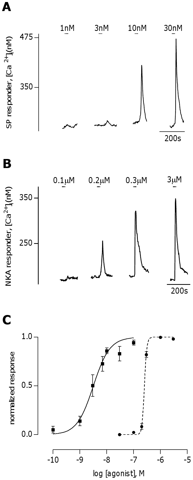
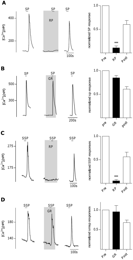
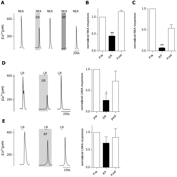
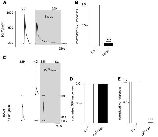
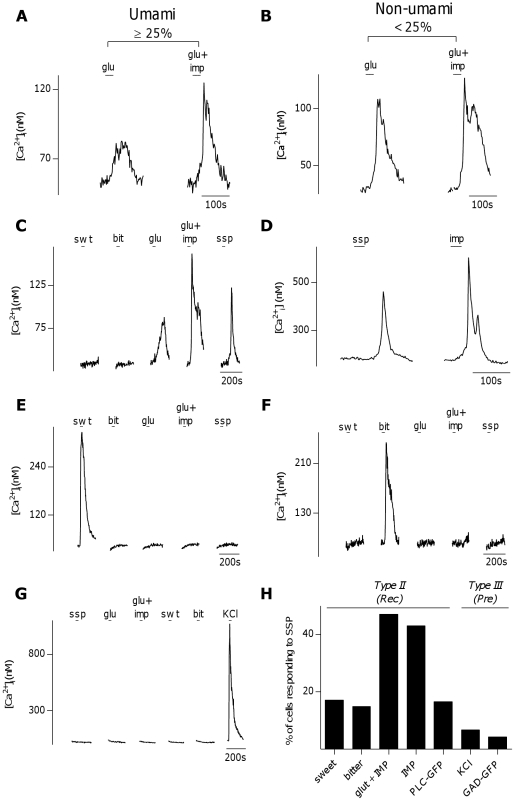
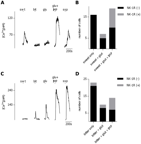
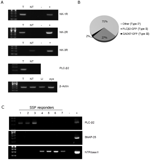
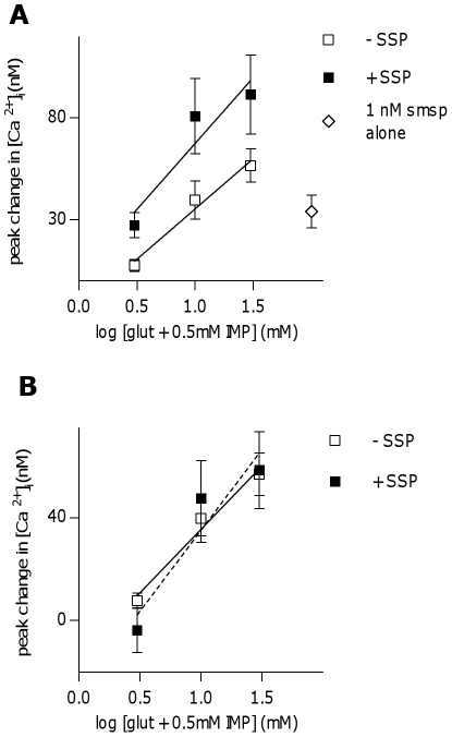
The tachykinins substance P (SP) and neurokinin A (NKA) are present in nociceptive sensory fibers expressing transient receptor potential cation channel, subfamily V, member 1 (TRPV1). These fibers are found extensively in and around the taste buds of several species. Tachykinins are released from nociceptive fibers by irritants such as capsaicin, the active compound found in chili peppers commonly associated with the sensation of spiciness. Using real-time Ca(2+)-imaging on isolated taste cells, it was observed that SP induces Ca(2+) -responses in a subset of taste cells at concentrations in the low nanomolar range. These responses were reversibly inhibited by blocking the SP receptor NK-1R. NKA also induced Ca(2+)-responses in a subset of taste cells, but only at concentrations in the high nanomolar range. These responses were only partially inhibited by blocking the NKA receptor NK-2R, and were also inhibited by blocking NK-1R indicating that NKA is only active in taste cells at concentrations that activate both receptors. In addition, it was determined that tachykinin signaling in taste cells requires Ca(2+)-release from endoplasmic reticulum stores. RT-PCR analysis further confirmed that mouse taste buds express NK-1R and NK-2R. Using Ca(2+)-imaging and single cell RT-PCR, it was determined that the majority of tachykinin-responsive taste cells were Type I (Glial-like) and umami-responsive Type II (Receptor) cells. Importantly, stimulating NK-1R had an additive effect on Ca(2+) responses evoked by umami stimuli in Type II (Receptor) cells. This data indicates that tachykinin release from nociceptive sensory fibers in and around taste buds may enhance umami and other taste modalities, providing a possible mechanism for the increased palatability of spicy foods.


Conflict of interest statement

Competing Interests: The author has declared that no competing interests exist.

Figures

Figure 1
Figure 1. Substance P and Neurokinin A induce Ca2+-responses in mouse taste cells.
A, Representative Ca2+ trace from a substance P (SP) responsive taste cell stimulated with 1,3, 10, and 30 nM SP. B, Representative Ca2+ trace from a neurokinin A (NKA) responsive taste cell stimulated with 0.1 ,0.2, 0.3, and 3 µM NKA. C, Dose response curves for SP (solid line, n = 33 cells) and NKA (dotted line, n = 37 cells) in isolated taste cells, normalized to the concentration of agonist that gives the highest Ca2+-response in a given cell. SP had an EC50 of 3.2 nM, while NKA had an EC50 of 256 nM.
Figure 2
Figure 2. Substance P-induced Ca2+-responses in mouse taste cells involve activation of the neurokinin 1 receptor.
A, Inhibition of Ca2+- responses to substance P (SP, 10 nM) in an isolated taste cell by the neurokinin 1 receptor (NK-1R) antagonist RP67580 (RP, 100 nM). Right panel: RP67580 reversibly inhibits SP-induced Ca2+-responses. Bars represent averaged normalized peak responses from 9 cells (***P<0.0001, repeated measures ANOVA). B, Example trace from isolated taste cell showing no inhibition of Ca2+-responses to SP (10 nM) by the neurokinin 2 receptor (NK-2R) antagonist GR159897 (GR,1 µM). Right panel: GR159897 does not inhibit SP-induced Ca2+- responses. Bars represent averaged normalized peak responses from 9 cells. C, Inhibition of Ca2+- responses to NK-1R-selective agonist [Sar9,Met(O2)11]-Substance P (SSP, 3 nM) in an isolated taste cell by RP67580 (RP, 30 nM). Right panel: RP67580 reversibly inhibits SSP-induced Ca2+-responses. Bars represent averaged normalized peak responses from 4 cells (***P<0.0001, repeated measures ANOVA). D, Example trace from isolated taste cell showing no inhibition of Ca2+-responses to SSP (3 nM) by GR159897 (1 µM). Right panel: GR159897 does not inhibit SSP-induced Ca2+ responses. Bars represent averaged normalized peak responses from 8 cells.
Figure 3
Figure 3. Neurokinin A-induced Ca2+-responses in mouse taste cells involve activation of both the neurokinin 1 and neurokinin 2 receptors.
A, Inhibition of Ca2+- responses to neurokinin A (NKA, 300 nM) in an isolated taste cell by the neurokinin 2 receptor (NK-2R) antagonist GR159897 (GR,1 µM) and the neurokinin 1 receptor (NK-1R) antagonist RP67580 (RP, 1 µM). B, GR159897 partially and reversibly inhibits NKA-induced Ca2+- responses. Bars represent averaged normalized peak responses from 30 cells (***P<0.0001, repeated measures ANOVA). C, RP67580 reversibly inhibits NKA-induced Ca2+-responses. Bars represent averaged normalized peak responses from 7 cells (***P<0.0001, repeated measures ANOVA). D, Inhibition of Ca2+- responses to [Lys5,MeLeu9,Nle10]-NKA(4–10) (LN, 100 nM) in an isolated taste cell by GR159897 (GR, 300 nM). Right panel: GR159897 reversibly inhibits LN-induced Ca2+- responses. Bars represent averaged normalized peak responses from 7 cells (*P<0.05, repeated measures ANOVA). E, Example trace from isolated taste cell showing no significant inhibition of Ca2+-responses to LN (100 nM) by RP67580 (1 µM). Right panel: RP67580 does not inhibit LN-induced Ca2+- responses. Bars represent averaged normalized peak responses from 7 cells.
Figure 4
Figure 4. NK-1R activation induces Ca2+ release from intracellular stores in taste cells.
A, Inhibition of Ca2+ responses to [Sar9,Met(O2)11]-Substance P (SSP, 10 nM) in an isolated taste cell after a 10 minute pre-treatment with the sarco-endoplasmic reticulum Ca2+-ATPase inhibitor thapsigargin (Thaps, 1 µM). B, Thapsigargin inhibits SSP-induced Ca2+-responses in taste cells. Bars represent averaged normalized peak responses from 10 cells (***P<0.0001, student's t-test). C, Representative Ca2+ traces from Type III (Presynaptic) and [Sar9,Met(O2)11]-Substance P (SSP)-responsive taste cells stimulated in both the presence and absence of extracellular Ca2+ in the same experimental run. The Type III (Presynaptic) cell (top trace) responded to 50 mM KCl only while extracellular Ca2+ was present, while the SSP-responsive cell (bottom trace) showed similar responses to 10 nM SSP in both the presence and absence of extracellular Ca2+. D, Ca2+ responses to 10 nM SSP in SSP-responsive taste cells were not significantly changed in the absence of extracellular Ca2+. Bars represent averaged normalized peak responses from 27 cells. E, Ca2+ responses to 50 mM KCl in Type III (Presynaptic) cells were eliminated in the absence of extracellular Ca2+. Bars represent averaged normalized peak responses from 11 cells (*** p<0.0001, Student's paired t-test).
Figure 5
Figure 5. Activation of neurokinin 1 receptors induces Ca2+ responses in a subset of Type II (Receptor) cells.
A, B, Physiological criteria for identifying umami Type II (Receptor) cells. A, Ca2+-responses to glutamate (30 mM) and glutamate+IMP (0.5 mM) in a taste cell. This cell showed a greater than 25% increase in peak response to glutamate in the presence of IMP, thus was defined as an umami cell. B. Another taste cell, which did not show a greater than 25% increase in peak response to glutamate in the presence of IMP. This cell was defined as a non-umami cell. C, An umami cell, characterized by increased sensitivity to glutamate (glu, 30 mM) in the presence of IMP (0.5 mM), also showed a Ca2+-response to [Sar9,Met(O2)11]-Substance P (ssp ,10 nM). A large number of umami cells (47%, 124/264 cells) responded to ssp. D, Similarly, sample trace of an umami cell, characterized by responsiveness to a high concentration of IMP alone (2.5 mM), which also responded to ssp (10 nM). E+F, sweet- (swt,E) or bitter- (bit, F) responsive cell that was unresponsive to bath-applied ssp, (10 nM). Only 6 of 35 (17.1%) sweet and 24 of 162 (14.8%) bitter taste cells responded to SSP. G, A Type III (Presynaptic) cell, characterized by Ca2+-influx through voltage gated calcium channels due to depolarization by KCl (50 mM), that was unresponsive to SSP (10 nM). Only 7 of 104 (6.7%) of physiologically identified Type III (Presynaptic) cells responded to SSP. H, Summary of the percent of physiologically identified Type II (Receptor) (Rec) and Type III (Presynaptic)(Pre) cells that responded to SSP.
Figure 6
Figure 6. A subset of sweet and bitter Type II (Receptor) cells respond to umami stimuli and express neurokinin 1 receptors.
A, Sweet- (swt) responsive taste cell that also showed Ca2+-responses to both umami stimuli (30 mM glu+0.5 mM IMP) and [Sar9,Met(O2)11]-Substance P (ssp,10 nM). B. Number of sweet only-, sweet- and glutamate-, and sweet-, glutamate- and IMP- responsive cells along with their respective expression of NK-1R as defined by responsiveness to ssp (10 nM). C, Bitter- (bit) responsive taste cell that also showed Ca2+-responses to both umami stimuli and SSP (10 nM). D, Number of bitter only-, bitter- and glutamate-, and bitter-, glutamate- and IMP- responsive cells along with their respective expression of NK-1R as defined by responsiveness to ssp (10 nM).
Figure 7
Figure 7. Neurokinin 1 receptor-expressing taste cells are Type I (Glial-like) cells and Type II (Receptor) cells.
A, The tachykinin receptors NK-1R and NK-2R are detectable by RT-PCR in mouse taste buds. RT-PCR for the tachykinin receptors NK-1R, NK-2R, and NK-3R in isolated taste buds (T), non-taste epithelium (NT), a negative control lacking template (−), and a positive control tissue (+): Large intestine (LI) for NK-1R and NK-2R, eye for NK-3R. PLC-β2 was used to validate distinct taste bud cDNA versus non-taste epithelium cDNA, and β-Actin was a positive control for quality of the template cDNAs. B, Identity of cells responding to 10 nM [Sar9,Met(O2)11]-Substance P (SSP) in experiments using isolated taste cells from PLCβ2-GFP and GAD67-GFP transgenic mice. ∼27% of SSP responders were PLCβ2-GFP (+) (dark grey), while only ∼2% were GAD67-GFP (+) (black), indicating that a large proportion (∼70%) of SSP-responders were of an unknown cell type, possibly Type I taste cells (light grey). C, RT-PCR for PLC-β2, SNAP-25, and NTPdase II in isolated taste cells that showed calcium responses to 10 nM SSP (1–7), a negative control lacking template (−), and whole isolated taste buds used as a positive control (+). Cells 1–3 expressed PLC-β2, but not SNAP-25, and NTPdase II, while cells 4–7 expressed NTPdase II, but not PLC-β2 or SNAP-25.
Figure 8
Figure 8. Stimulation of the neurokinin 1 receptor has an additive effect on umami responses in taste cells.
A, Peak Ca2+-responses to 3 concentrations of glutamate (3, 10, 30 mM)+0.5 mM IMP are significantly increased in the presence of 1 nM [Sar9,Met(O2)11]-Substance P (SSP, 1 nM) in SSP-responsive umami taste cells (p = 0.03682, linear regression analysis). Open squares represent averaged responses to glutamate+IMP alone, closed squares represent averaged responses to glutamate+IMP+1 nM SSP, while open diamond represents average response to 1 nM SSP alone. B, When peak response to SSP alone is subtracted from responses to glutamate+IMP+SSP in each cell (dotted line), there is no difference in responses as compared to glutamate+IMP alone (solid line).

Similar articles

Cited by

References

    1. Caterina MJ, Julius D. The vanilloid receptor: a molecular gateway to the pain pathway. Annu Rev Neurosci. 2001;24:487–517. - PubMed
    1. Caterina MJ, Schumacher MA, Tominaga M, Rosen TA, Levine JD, et al. The capsaicin receptor: a heat-activated ion channel in the pain pathway. Nature. 1997;389:816–824. - PubMed
    1. Pennefather JN, Lecci A, Candenas ML, Patak E, Pinto FM, et al. Tachykinins and tachykinin receptors: a growing family. Life Sci. 2004;74:1445–1463. - PubMed
    1. Moussaoui SM, Le Prado N, Bonici B, Faucher DC, Cuine F, et al. Distribution of neurokinin B in rat spinal cord and peripheral tissues: comparison with neurokinin A and substance P and effects of neonatal capsaicin treatment. Neuroscience. 1992;48:969–978. - PubMed
    1. Geppetti P, Nassini R, Materazzi S, Benemei S. The concept of neurogenic inflammation. BJU Int. 2008;101(Suppl 3):2–6. - PubMed

Publication types

MeSH terms