. 2012;7(2):e31850.
doi: 10.1371/journal.pone.0031850. Epub 2012 Feb 20.

iPLA2β overexpression in smooth muscle exacerbates angiotensin II-induced hypertension and vascular remodeling

Affiliations

iPLA2β overexpression in smooth muscle exacerbates angiotensin II-induced hypertension and vascular remodeling

Lindsay E Calderon et al. PLoS One. 2012.

Abstract

Objectives: Calcium independent group VIA phospholipase A(2) (iPLA(2)β) is up-regulated in vascular smooth muscle cells in some diseases, but whether the up-regulated iPLA(2)β affects vascular morphology and blood pressure is unknown. The current study addresses this question by evaluating the basal- and angiotensin II infusion-induced vascular remodeling and hypertension in smooth muscle specific iPLA(2)β transgenic (iPLA(2)β-Tg) mice.

Method and results: Blood pressure was monitored by radiotelemetry and vascular remodeling was assessed by morphologic analysis. We found that the angiotensin II-induced increase in diastolic pressure was significantly higher in iPLA(2)β-Tg than iPLA(2)β-Wt mice, whereas, the basal blood pressure was not significantly different. The media thickness and media∶lumen ratio of the mesenteric arteries were significantly increased in angiotensin II-infused iPLA(2)β-Tg mice. Analysis revealed no difference in vascular smooth muscle cell proliferation. In contrast, adenovirus-mediated iPLA(2)β overexpression in cultured vascular smooth muscle cells promoted angiotensin II-induced [(3)H]-leucine incorporation, indicating enhanced hypertrophy. Moreover, angiotensin II infusion-induced c-Jun phosphorylation in vascular smooth muscle cells overexpressing iPLA2β to higher levels, which was abolished by inhibition of 12/15 lipoxygenase. In addition, we found that angiotensin II up-regulated the endogenous iPLA(2)β protein in-vitro and in-vivo.

Conclusion: The present study reports that iPLA(2)β up-regulation exacerbates angiotensin II-induced vascular smooth muscle cell hypertrophy, vascular remodeling and hypertension via the 12/15 lipoxygenase and c-Jun pathways.


Conflict of interest statement

Competing Interests: The authors have declared that no competing interests exist.

Figures

Figure 1
Figure 1. Enhanced diastolic blood pressure increase in iPLA2β-Tg mice in response to Ang II infusion.
Blood pressure was measured in 13-wk old male, iPLA2β-Wt and iPLA2β-Tg mice by radiotelemetry. 24 h averages of systolic (A), diastolic (B), and mean arterial pressure (C), heart rate (D), pulse pressure (E), and locomotor activity (F) were collected prior to and during Ang II infusion (500 ng/kg/min, 14 days). n = 8; *: p<0.05 by two-way ANOVA with repeated measures.
Figure 2
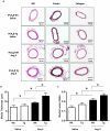
Figure 2. Exacerbated mesenteric artery remodeling in iPLA2β-Tg mice in response to Ang II infusion.
Vascular remodeling was analyzed in embedded sections of the 2nd order branch of mesenteric arteries from the iPLA2β-Wt and iPLA2β-Tg mice infused with saline or Ang II (500 ng/kg/min, 14 days). Representative images of vessel sections stained with HE, elastin or collagen (A). The media thickness (B) and media∶lumen ratio (C) were quantified. n = 8, *: p<0.05, **: p<0.01, ***: p<0.001 by one-way ANOVA.
Figure 3
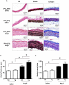
Figure 3. Exacerbated thoracic aorta remodeling in iPLA2β-Tg mice in response to Ang II infusion.
Vascular remodeling was analyzed in the thoracic aorta of the iPLA2β-Wt and iPLA2β-Tg infused with Ang II (500 ng/kg/min) or saline. Representative images of vessel sections stained with HE, elastin or collagen (A). The media thickness (B) and media∶lumen ratio (C) were quantified. n = 8, **: p<0.01, ***: p<0.001 by one way ANOVA.
Figure 4
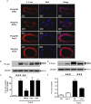
Figure 4. Lack of difference in cell proliferation between iPLA2β-Tg and iPLA2β-Wt mice by Ang II infusion.
Mice were infused with saline or Ang II (500 ng/kg/min, 14 days) and thoracic aortas were isolated and sectioned. (A) are representative images of the sections stained with anti-PCNA antibody. (B) and (C) are summaries of the cell number analysis using HE stained sections. n = 8.
Figure 5
Figure 5. iPLA2β overexpression promotes Ang II-induced [3H]-leucine incorporation in VSMC via the 12/15 lipoxygenase pathway.
[3H]-leucine incorporation was quantified by liquid scintillation spectroscopy in cultured iPLA2β-Wt VSMCs treated with Ang II (100 nM, 24 h) or saline after (A) Ad-iPLA2β (500 MOI) and Ad-tet-on (2000 MOI), and/or doxycycline (1 µg/ml); (B) saline or inhibitors of lipoxygenase (NDGA, 30 µM), or cyclooxygenase (Indomethacin, 50 µM) or P450 cytochrome C (ODA, 10 µM). (C) Lox−/− or Wt VSMCs stimulated with Ang II or saline. n = 3–4, *: p<0.05, **: p<0.01, ***: p<0.001 one-way ANOVA.
Figure 6
Figure 6. iPLA2β overexpression promotes Ang II-induced c-Jun phosphorylation via the 12/15 lipoxygenase pathway.
(A) iPLA2β-Wt and iPLA2β-Tg mice were infused with Ang II (1000 ng/kg/min, 14 days) or saline and 2nd order branch of mesenteric arteries VSMC were isolated and embedded in OCT. Vessel sections were stained with anti-p-c-Jun antibody and DAPI. (B) and (D) are representative western blots, (C) and (E) are quantifications of the blots, of c-Jun phosphorylation in aortic VSMCs after the same treatments as in Fig. 3 B and C. n = 3–4, *:p<0.05, **: p<0.01, ***: p<0.001 one-way ANOVA.
Figure 7
Figure 7. Lack of effects of inhibiting arachidonic acid metabolism on Ang II induced p38 MAPK activation.
VSMC were isolated from the aortas of iPLA2β-Wt mice and incubated with inhibitors of lipoxygenase (NDGA, 30 µM), cyclooxygenase (Indomethacin, 50 µM) or P450 cytochrome C (ODA, 10 µM) and stimulated with Ang II (100 nM) or saline for 1 h. Then the total- or phosphorylated p38 MAPK was determined by Western Blot. n = 3–4.
Figure 8
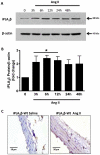
Figure 8. Ang II enhances iPLA2β protein expression in cultured VSMC and in-vivo.
24 h serum-deprived rat aortic VSMC were stimulated with Ang II (100 nM) for various time intervals as indicated. iPLA2β protein abundance was determined by Western Blot (A and B). Representative immunohistochemical images of iPLA2β protein staining in C57/B6 thoracic aorta from iPLA2β-Wt and iPLA2β-Tg mice infused with saline or Ang II (500 ng/kg/min, 14 days) (C). n = 4–5; *: p<0.05 by one-way ANOVA analysis.

Similar articles

Cited by

References

    1. Murakami M, Taketomi Y, Miki Y, Sato H, Hirabayashi T, et al. Recent progress in phospholipase A research: from cells to animals to humans. Progress in lipid research. 2011;50:152–192. - PubMed
    1. Khateeb S, Flusser H, Ofir R, Shelef I, Narkis G, et al. PLA2G6 mutation underlies infantile neuroaxonal dystrophy. Am J Hum Genet. 2006;79:942–948. - PMC - PubMed
    1. Morgan NV, Westaway SK, Morton JE, Gregory A, Gissen P, et al. PLA2G6, encoding a phospholipase A2, is mutated in neurodegenerative disorders with high brain iron. Nat Genet. 2006;38:752–754. - PMC - PubMed
    1. Ford DA, Hazen SL, Saffitz JE, Gross RW. The rapid and reversible activation of a calcium-independent plasmalogen-selective phospholipase A2 during myocardial ischemia. J Clin Invest. 1991;88:331–335. - PMC - PubMed
    1. Mancuso DJ, Abendschein DR, Jenkins CM, Han X, Saffitz JE, et al. Cardiac ischemia activates calcium-independent phospholipase A2beta, precipitating ventricular tachyarrhythmias in transgenic mice: rescue of the lethal electrophysiologic phenotype by mechanism-based inhibition. The Journal of biological chemistry. 2003;278:22231–22236. - PubMed

Publication types

MeSH terms