. 2012;7(2):e31852.
doi: 10.1371/journal.pone.0031852. Epub 2012 Feb 21.

Low-resolution molecular models reveal the oligomeric state of the PPAR and the conformational organization of its domains in solution

Affiliations

Low-resolution molecular models reveal the oligomeric state of the PPAR and the conformational organization of its domains in solution

Amanda Bernardes et al. PLoS One. 2012.

Abstract

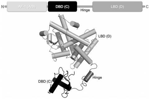
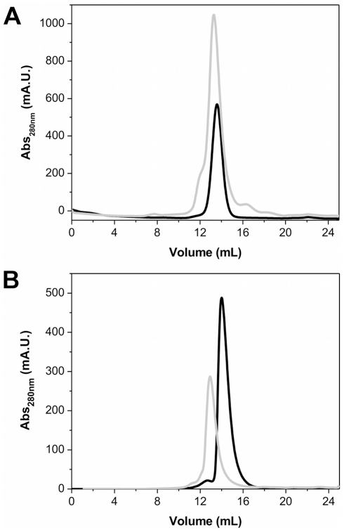
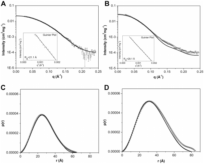
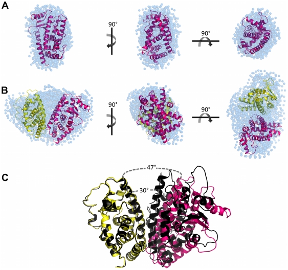
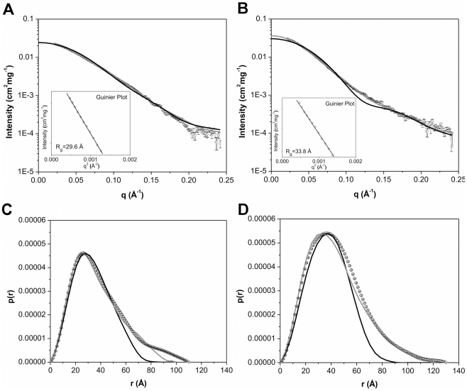
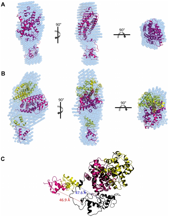
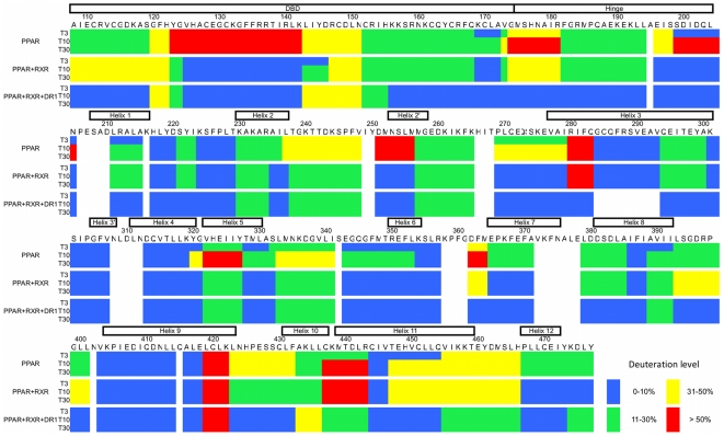
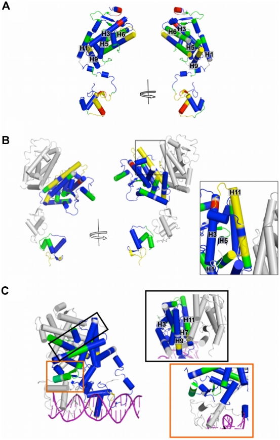
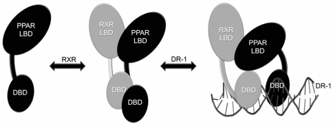
The peroxisome proliferator-activated receptors (PPARs) regulate genes involved in lipid and carbohydrate metabolism, and are targets of drugs approved for human use. Whereas the crystallographic structure of the complex of full length PPARγ and RXRα is known, structural alterations induced by heterodimer formation and DNA contacts are not well understood. Herein, we report a small-angle X-ray scattering analysis of the oligomeric state of hPPARγ alone and in the presence of retinoid X receptor (RXR). The results reveal that, in contrast with other studied nuclear receptors, which predominantly form dimers in solution, hPPARγ remains in the monomeric form by itself but forms heterodimers with hRXRα. The low-resolution models of hPPARγ/RXRα complexes predict significant changes in opening angle between heterodimerization partners (LBD) and extended and asymmetric shape of the dimer (LBD-DBD) as compared with X-ray structure of the full-length receptor bound to DNA. These differences between our SAXS models and the high-resolution crystallographic structure might suggest that there are different conformations of functional heterodimer complex in solution. Accordingly, hydrogen/deuterium exchange experiments reveal that the heterodimer binding to DNA promotes more compact and less solvent-accessible conformation of the receptor complex.


Conflict of interest statement

Competing Interests: The authors have declared that no competing interests exist.

Figures

Figure 1
Figure 1. Structural organization of nuclear receptors functional domains.
A) Bar representation of nuclear receptors domains. B) Cartoon of crystallographic structure of intact PPARγ+RXRα+DR-1 complex (PDB 3DZU). The N-terminal region (A/B) represented by a light gray bar is absent in the structure because of it high flexibility. The conserved C region, which corresponds to the DBD, is given in black; the LBD, or region E, is shown in gray; and located between C and E domains, the hinge given here in dark gray.
Figure 2
Figure 2. Size exclusion chromatography profile showing the difference in the elution pattern of monomer and heterodimer proteins.
A) hPPARγ LBD and hPPARγ/hRXRα LBD and B) hPPARγ DBD-LBD and hPPARγ/hRXRα DBD-LBD. The SEC were performed on a Superdex 75 columm equilibrated with 20 mM Hepes-Na buffer (pH 8.0), 3 mM dithiothreitol, 200 mM NaCl, and 5% glycerol. Monomers are given in black solid lines and heterodimers in gray lines.
Figure 3
Figure 3. Small-angle X-ray scattering curves for LBD proteins construction.
A) hPPARγ LBDs 3.0 mg/mL and B) hPPARγ/hRXRα LBD heterodimers at 3.0 mg/mL. Experimental data (open black circles with errors bars), simulated curves corresponding to the high-resolution model obtained by the use of the PDB id 1FM6 (black solid line) and the rigid body model (gray line). Inset: Guinier plot. The distance distribution function from C) the hPPARγ LBD and D) the hPPARγ/hRXRα LBD. Experimental data (open black circles with errors bars), the high-resolution model (black solid line) and the rigid body model (gray line).
Figure 4
Figure 4. SAXS models for LBD proteins construction.
Three orthogonal views of the SAXS ab initio models for A) hPPARγ LBD, obtained by Gasbor (shaded spheres), superposed to the hPPARγ LBD monomeric part of the high-resolution model PDB id 1FM6 (cartoon) and B) hPPARγ/hRXRα LBD heterodimer, obtained by Gasbor (shaded spheres), superposed to the rigid body model from PDB id 1FM6 (cartoon). C) Superposition of the rigid body model with the crystallographic structure (PDB id 1FM6) showing the opening angle imposed on the rigid body model being larger than the crystallographic structure. hPPARγ LBD (pink), hRXRα LBD (yellow), crystallographic structure of heterodimer (black) and DAM (blue).
Figure 5
Figure 5. Small-angle X-ray scattering curves for DBD-LBD proteins construction.
A) hPPARγ DBD-LBD and B) hPPARγ/hRXRα DBD-LBD, both at 3.0 mg/mL. Experimental data (open black circles with errors bars), simulated curves corresponding to the high-resolution model obtained by the use of the PPARγ monomer from the PDB id 3DZU (black solid line) and the rigid body model (gray line). Inset: Guinier plot. Distance distribution function from C) the hPPARγ DBD-LBD and D) the hPPARγ/hRXRα DBD-LBD. Experimental data (open black circles with errors bars), the high-resolution model (black solid line) and the rigid body model (gray line).
Figure 6
Figure 6. SAXS Models for LBD proteins construction.
Three orthogonal views of the SAXS ab initio envelope for A) hPPARγ DBD-LBD, obtained by Dammin package, superposed to the monomer rigid body model from PDB id 3DZU and B) hPPARγ/hRXRα DBD-LBD heterodimer, obtained by Dammin package, superposed to the heterodimer rigid body model from PDB id 3DZU. C) Superposition of the rigid body model with the crystallographic structure (PDB id 3DZU) showing the differences between the DBDs positions of the rigid body model and the crystallographic structure. The DBDs was translated and the distance between the initial and final position of them is represented by red dotted line for DBDs hPPARγ and blue dotted line for DBDs of hRXRα. In pink is hPPARγ LBD and yellow is hRXRα LBD of rigid body model; heterodimer crystallographic structure (dark pink and dark yellow) (PDB id 3DZU) and DAM (blue).
Figure 7
Figure 7. Deuteration level of PPAR monomer and in complex with RXR and DR-1.
Deuteration level according with the PPAR sequence, showing the dynamic features of the protein in solution. Each block of three lines represent one protein sample (PPAR monomer, PPAR+RXR complex in absence and in presence of DR-1) in three different deuterium incubation time (3, 10 and 30 minutes). The sequence colored according to H/D-Ex data, considering blue as 0–10%, green 11–30%, yellow 31–50% and red >50% of D2O incorporation.
Figure 8
Figure 8. PPAR DBD-LBD models colored according to H/D Ex-data.
Protections and solvent exposure are colored according deuteration level, from blue (0–10% D2O incorporation), green (11–30% D2O incorporation), yellow (31–50% D2O incorporation) to red (more that 50% of D20 incorporation). A) hPPARγ monomer; B) hPPARγ/hRXRα heterodimer. The box shows in details the dimerization interface, with H10-11 being not very strongly protected (yellow - 11 to 49% D2O incorporation). C) hPPARγ/hRXRα+DR1 complex, the boxes show dimerization interface (top box, framed in black), which presents H10-11 and H7 more protected than that in hPPARγ/hRXRα heterodimer alone; and the third heterodimerization interface (bottom box – orange) indicating higher degree of protection.
Figure 9
Figure 9. Cartoon schematically representing the mechanism of heterodimerization and binding to the DNA.
When the PPAR is activated, it recruits RXR, forming an intermediary heterodimer, which has the LBDs and DBDs domains in extended and open conformation. Following to DNA binding, the PPAR/RXR heterodimers suffer additional conformational changes, becoming more condensed and less solvent-exposed.

Similar articles

Cited by

References

    1. Chiarelli F, Di Marzio D. Peroxisome proliferator-activated receptor-gamma agonists and diabetes: current evidence and future perspectives. Vasc Health Risk Manag. 2008;4:297–304. - PMC - PubMed
    1. Michalik L, Auwerx J, Berger J, Chatterjee V, Glass C, et al. International Union of Pharmacology. LXI. Peroxisome proliferator-activated receptors. Pharmacol Rev. 2006;58:726–741. - PubMed
    1. Szanto A, Nagy L. The many faces of PPARgamma: anti-inflammatory by any means? Immunobiology. 2008;213:789–803. - PubMed
    1. Aranda A, Pascual A. Nuclear hormone receptors and gene expression. Physiol Rev. 2001;81:1269–1304. - PubMed
    1. Desvergne B, Wahli W. Peroxisome proliferator-activated receptors: nuclear control of metabolism. Endocr Rev. 1999;20:649–688. - PubMed

Publication types

MeSH terms