

The dispanins: a novel gene family of ancient origin that contains 14 human members
- PMID: 22363774
- PMCID: PMC3282796
- DOI: 10.1371/journal.pone.0031961
The dispanins: a novel gene family of ancient origin that contains 14 human members
Abstract
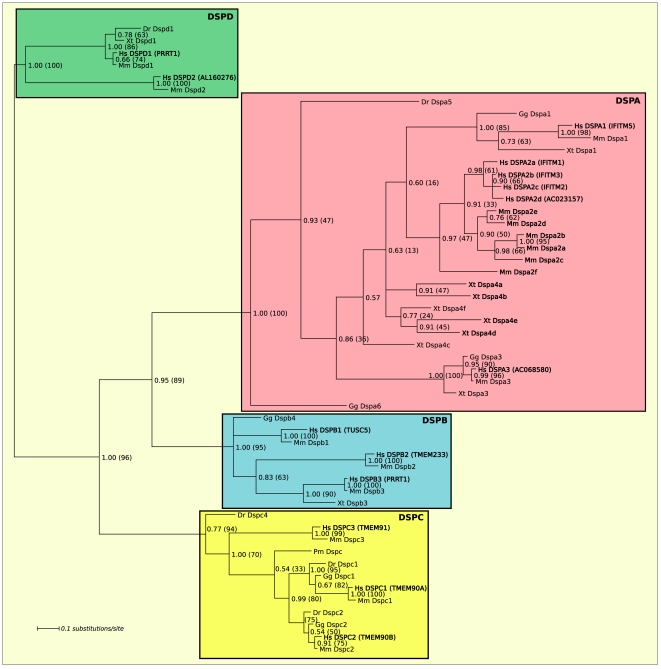
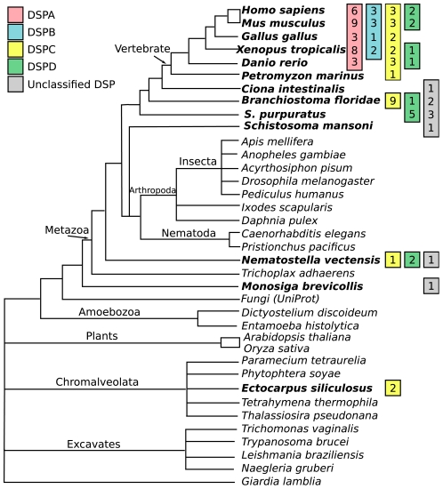
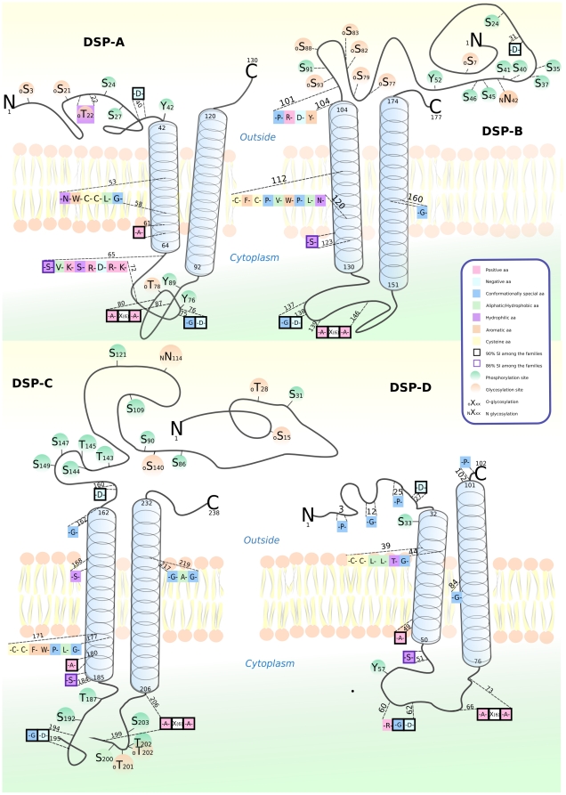
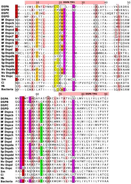
The Interferon induced transmembrane proteins (IFITM) are a family of transmembrane proteins that is known to inhibit cell invasion of viruses such as HIV-1 and influenza. We show that the IFITM genes are a subfamily in a larger family of transmembrane (TM) proteins that we call Dispanins, which refers to a common 2TM structure. We mined the Dispanins in 36 eukaryotic species, covering all major eukaryotic groups, and investigated their evolutionary history using Bayesian and maximum likelihood approaches to infer a phylogenetic tree. We identified ten human genes that together with the known IFITM genes form the Dispanin family. We show that the Dispanins first emerged in eukaryotes in a common ancestor of choanoflagellates and metazoa, and that the family later expanded in vertebrates where it forms four subfamilies (A-D). Interestingly, we also find that the family is found in several different phyla of bacteria and propose that it was horizontally transferred to eukaryotes from bacteria in the common ancestor of choanoflagellates and metazoa. The bacterial and eukaryotic sequences have a considerably conserved protein structure. In conclusion, we introduce a novel family, the Dispanins, together with a nomenclature based on the evolutionary origin.
Conflict of interest statement
Figures
References
-
- Rask-Andersen M, Almen MS, Schioth HB. Trends in the exploitation of novel drug targets. Nat Rev Drug Discov. 2011;10:579–590. - PubMed
-
- Fredriksson R, Lagerstrom MC, Lundin LG, Schioth HB. The G-protein-coupled receptors in the human genome form five main families. Phylogenetic analysis, paralogon groups, and fingerprints. Mol Pharmacol. 2003;63:1256–1272. - PubMed
-
- Lagerstrom MC, Schioth HB. Structural diversity of G protein-coupled receptors and significance for drug discovery. Nat Rev Drug Discov. 2008;7:339–357. - PubMed
-
- Fredriksson R, Nordstrom KJ, Stephansson O, Hagglund MG, Schioth HB. The solute carrier (SLC) complement of the human genome: phylogenetic classification reveals four major families. FEBS Lett. 2008;582:3811–3816. - PubMed
Publication types
MeSH terms
Substances
LinkOut - more resources
Full Text Sources
Molecular Biology Databases
Miscellaneous
