. 2011:2011:275782.
doi: 10.5402/2011/275782. Epub 2011 Jul 12.

Culture models for studying thyroid biology and disorders

Affiliations

Culture models for studying thyroid biology and disorders

Shuji Toda et al. ISRN Endocrinol. 2011.

Abstract

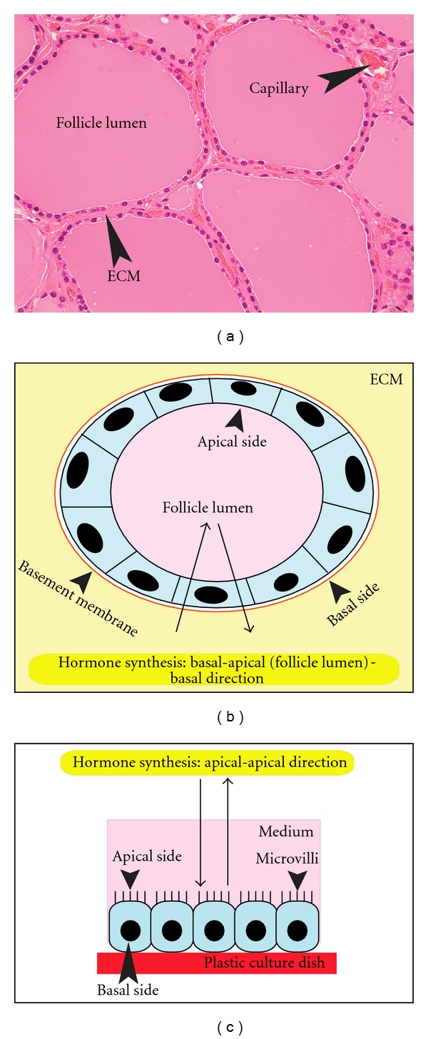
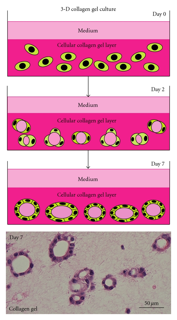
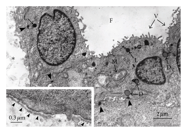
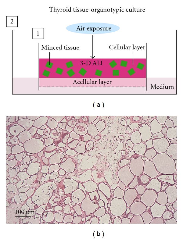
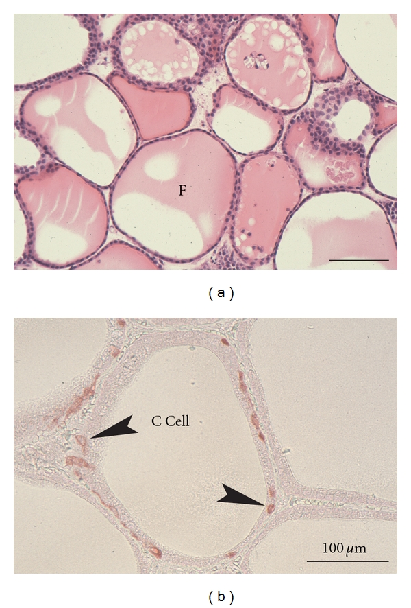
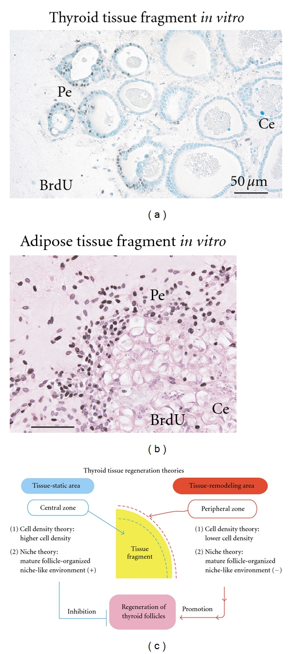
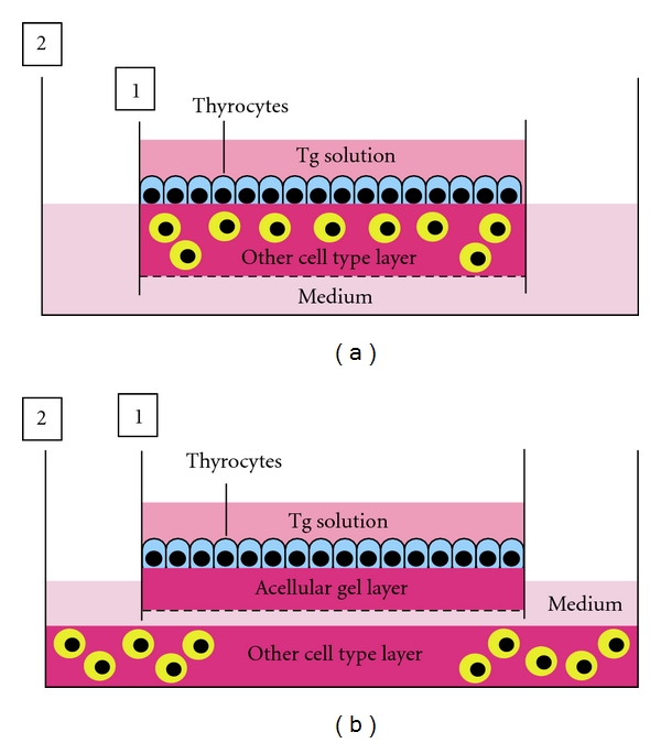
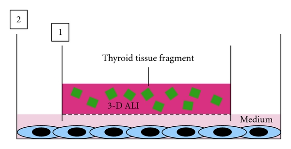
The thyroid is composed of thyroid follicles supported by extracellular matrix, capillary network, and stromal cell types such as fibroblasts. The follicles consist of thyrocytes and C cells. In this microenvironment, thyrocytes are highly integrated in their specific structural and functional polarization, but monolayer and floating cultures cannot allow thyrocytes to organize the follicles with such polarity. In contrast, three-dimensional (3-D) collagen gel culture enables thyrocytes to form 3-D follicles with normal polarity. However, these systems never reconstruct the follicles consisting of both thyrocytes and C cells. Thyroid tissue-organotypic culture retains 3-D follicles with both thyrocytes and C cells. To create more appropriate experimental models, we here characterize four culture systems above and then introduce the models for studying thyroid biology and disorders. Finally, we propose a new approach to the cell type-specific culture systems on the basis of in vivo microenvironments of various cell types.


Figures

Figure 1
Figure 1
(a) Histology of the thyroid gland, comprised of colloid-filled thyroid follicles, which are supported by extracellular matrix (ECM) and a capillary network. (b) A schema of a thyroid follicle in vivo and in 3-D collagen gel culture. The follicle is embedded in ECM. The component thyrocytes of the follicle show a specific cellular polarity: the apical side with microvilli faces the follicle lumen, and the basal pole with basal lamina faces the ECM. Thyrocytes undergo thyroid hormone synthesis and release in a basal-apical (follicle lumen)-basal direction. (c) A schema of thyrocytes in a monolayer culture system. The cells have a cellular polarity: the apical pole with microvilli faces culture medium, and the basal side without basal lamina faces the plastic surface. Thyrocytes in such an environment carry out thyroid hormone synthesis and release only in an apical-apical direction.
Figure 2
Figure 2
Scheme of 3-D collagen gel culture system and histology of thyroid follicles reconstructed in its system. Thyrocytes embedded in 3-D type I collagen gel (day 0) reconstruct small thyroid follicles (day 2). Thyroid follicles grow larger through proliferation of component thyrocytes (day 7). The lowest panel shows the histology of reconstructed thyroid follicles. Hematoxylin and eosin (H&E) stain.
Figure 3
Figure 3
Fine structures of thyroid follicles reconstructed in 3-D collagen gel culture. The thyrocytes have numerous microvilli (V) at the apical surface of the follicle lumen (F) and form basal lamina (small arrowheads in inset) at the basal side which makes contact with collagen gel (∗). The cells have lysosomes (L), colloid droplets (D), and mitochondria (M). Junctional complexes (large arrowheads) are organized at the contact points of the cells.
Figure 4
Figure 4
Scheme of thyroid tissue-organotypic culture system and histology of thyroid tissue maintained in its system. (a) Minced tissues embedded in type I collagen gel (cellular layer) are placed on the acellular gel (acellular layer) in inner dish (1). The inner dish (1) is put in the outer dish (2) with culture medium. In this way, the tissues in cellular layer are localized under air exposure-induced oxygenation (3-D ALI). The tissues are kept in moist and fed by culture medium that percolates by capillary action from the medium-containing outer dish, through the acellular layer and into the cellular layer. (b) Many viable thyroid follicles are maintained at 3 months in this culture system. H&E stain.
Figure 5
Figure 5
Histology of thyroid tissues (a) and immunohistochemistry for calcitonin (b) in thyroid tissue-organotypic culture. (a) At 40 days in culture, viable thyroid follicles enclosed by thyrocytes contain colloid substance in their lumens (F). H&E stain. (b) At 30 days in culture, thyroid follicles consisting of both thyrocytes and calcitonin-positive C cells (arrowheads) are clearly maintained.
Figure 6
Figure 6
Immunohistochemistry with the growth marker BrdU in the organotypic cultures of thyroid (a) and adipose (b) tissue fragments, and scheme of thyroid tissue regeneration theories (c). (a) and (b) Intranuclear BrdU uptakes of thyrocytes and adipose cell types at peripheral zone (Pe) of thyroid (a) and adipose tissue fragments (b) are extensively greater than those at the central zone (Ce). Pe: peripheral zone. Ce: central zone. (c) Two theories regarding the mechanism of thyroid follicle regeneration that occurs specifically at peripheral zone of the tissue fragments. In regard to “cell density theory”, central zone of thyroid tissue fragments is characterized by higher cell density, whereas the peripheral zone is characterized by lower cell density. In general, increased cell density in a microenvironment inhibits the regeneration and growth of cells that are subjected to contact the inhibition of cell growth. Namely, central zone is tissue-static area with cell growth inactivation, while peripheral zone is tissue-remodeling area with cell growth activation. Thus, lower cell density of the peripheral zone may contribute to active development of thyroid follicles. In regard to “niche theory”, central zone concentrated by mature thyroid follicles may be subjected to mature thyroid follicle-organized niche-like environment, whereas peripheral zone with sparse population of the mature follicles may lose the environment. In general, a niche environment for stem cell types maintains their resting state. Thus, the niche-like environment formed by the mature follicles may inhibit regeneration of the follicles at the center, while its loss at the peripheral zone may contribute to their regeneration.
Figure 7
Figure 7
Models for analyzing thyrocyte-other cell type interaction. (a) Tg solution is overlaid on the surface of thyrocytes monolayer-cultured on other cell type-embedded collagen gel layer in an inner dish (1) with nitrocellulose membrane in its bottom. The inner dish is placed in a larger outer dish (2), and then culture medium is added to the outer dish. (b) To easily analyze the protein, mRNA, and DNA expression of both thyrocytes and other cell types under their interactions, the inner dish, in which Tg solution-exposing thyrocytes are cultured on acellular collagen gel layer, is placed in the outer dish where other cell types are cultured in a monolayer or 3-D collagen gel.
Figure 8
Figure 8
A model for analyzing thyroid tissue-other cell type interaction. Thyroid tissue fragments are embedded in ALI-treated collagen gel in an inner dish (1) with a nitrocellulose membrane. The inner dish is placed in a larger outer dish (1) in which each of other cell types is already cultured in monolayer or 3-D collagen gel. Then, culture medium is added to the outer dish.
Figure 9
Figure 9
Scheme of a new classification of culture system. (a) Liquid-rich monolayer culture system. The usual monolayer culture under a submerged condition with enough medium is suitable for culturing the surface-lining cell types of endothelial cells, ependymocytes, renal tubular cells, and so on. (b) 2-D ALI culture system is useful for culturing the surface-lining cell types of epidermis, cornea, and respiratory and digestive tracts. (c) 3-D ALI culture is suitable for culturing parenchymal and stromal cell types of solid organs.

References

    1. Fawcett DW. The thyroid gland. In: Fawcett DW, Raviola E, editors. Textbook of Histology. New York, NY, USA: Chapman & Hall; 1994. pp. 490–502.
    1. Pulvertaft RJV, Davies JR, Weiss L, Wilkinson JHJ. Studies on tissue cultures of human pathological thyroids. The Journal of Pathology and Bacteriology. 1959;77(1):19–32. - PubMed
    1. Takasu N. Primary culture of thyroid cells. In: Griffiths JB, Doyle A, Newell DG, editors. Cell and Tissue Culture: Laboratory Procedures. 17B (1) Chichester, UK: John Wiley & Sons; 1996. pp. 1–13.
    1. Toda S, Sugihara H. Primary culture of the thyroid: three-dimensional culture using extracellular matrix. In: Griffiths JB, Doyle A, Newell DG, editors. Cell and Tissue Culture: Laboratory Procedures. 17B (2) Chichester, UK: John Wiley & Sons; 1996. pp. 1–12.
    1. Toda S, Koike N, Sugihara H. Thyrocyte integration, and thyroid folliculogenesis and tissue regeneration: perspective for thyroid tissue engineering. Pathology International. 2001;51(6):403–417. - PubMed

LinkOut - more resources