Review
. 2012 Mar;14(1):31-45.
doi: 10.1016/j.jvc.2011.11.003. Epub 2012 Feb 24.

Differentiating the aging of the mitral valve from human and canine myxomatous degeneration

Affiliations
Review

Differentiating the aging of the mitral valve from human and canine myxomatous degeneration

Patrick S Connell et al. J Vet Cardiol. 2012 Mar.

Abstract

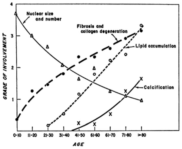
During the course of both canine and human aging, the mitral valve remodels in generally predictable ways. The connection between these aging changes and the morbidity and mortality that accompany pathologic conditions has not been made clear. By exploring work that has investigated the specific valvular changes in both age and disease, with respect to the cells and the extracellular matrix found within the mitral valve, heretofore unexplored connections between age and myxomatous valve disease can be found. This review addresses several studies that have been conducted to explore such age and disease related changes in extracellular matrix, valvular endothelial and interstitial cells, and valve innervation, and also reviews attempts to correlate aging and myxomatous disease. Such connections can highlight avenues for future research and help provide insight as to when an individual diverts from an aging pattern into a diseased pathway. Recognizing these patterns and opportunities could result in earlier intervention and the hope of reduced morbidity and mortality for patients.


Figures

Figure 1
Figure 1
(A) Top (atrial) view of the mitral valve in the closed (left) and open (right) position. (B) Bottom (ventricular) view of the mitral valve, cut open at the commissures to display the posterior (left) and anterior (right) leaflets. Dotted white line demarcates leaflet center from free edge. Chordae may have basal or marginal insertion into leaflet. Scale bar = 1cm. Reprinted from Grande-Allen KJ, Calabro A, Gupta V, Wight TN, Hascall VC, Vesely I. Glycosaminoglycans and proteoglycans in normal mitral valve leaflets and chordae: association with regions of tensile and compressive loading. Glycobiology. 2004;14(7):621–33 with permission from Oxford Journals.
Figure 2
Figure 2
Changes in mitral valve with age averaged across 200 mitral valve samples. Trends include decreasing cellularity and increasing fibrosis and collagen degradation, lipid accumulation and calcification. Reprinted from Sell, S., Scully, R. E. (1965). Aging changes in the aortic and mitral valves: histologic and histochemical studies, with observations on the pathogenesis of calcific aortic stenosis and calcification of the mitral annulus. Am J Pathol, 46 (3), 345–365 with permission from Elsevier.
Figure 3
Figure 3
Proportion of glycosaminoglycans (GAGs) in the MVAC and MVF of 6-week-old, 6-month-old, and 6-year old porcine valves, as calculated from FACE. Statistically significant differences (p < 0.05) are labeled † (significant differences in MVAC and MVF at a given age) or * (significant differences across ages in a given region (MVAC or MVF)). α represents a lower p-value (p < 0.03) for one set of comparisons (I4S) between ages in a given region (MVF). Significance was not displayed for averaged data (i.e., difference between MVF and MVAC averaged across all ages). MVF = mitral valve (MV) free edge; MVAC = MV anterior center; G0S = unsulfated glucuronate; G4S = 4-sulfated glucuronate; G6S = 6-sulfated glucuronate; I4S = 4-sulfated iduronate; I6S = 6-sulfated iduronate; XS = di- and tri-sulfated glucuronate / iduronate. Only GAGs that comprise >10% are labeled. Modified from Stephens EH, Chu CK, Grande-Allen KJ. Valve proteoglycan content and glycosaminoglycan fine structure are unique to microstructure, mechanical load and age: relevance to an age-specific tissue-engineered heart valve. Acta Biomaterial. 2008;4(5):1148–1160 with permission from Elsevier.
Figure 4
Figure 4
Movat-stained circumferential sections of the MVAC and MVF of 6-week-old, 6-month-old, and 6-year-old porcine valves. Both high magnification (column 1) and low magnification (columns 2 – 4) are shown. All images within a magnification type (high vs. low) are the same magnification to allow comparison of leaflet and layer thickness across age and leaflet region. Movat pentachrome staining preferentially stains different ECM elements certain colors (yellow = aligned collagen, black = elastic fibers, and green/blue = PGs / GAGs). Comparing regions, the MVAC fibrosa comprised a larger proportion of the valve than in the MVF. Across ages (regardless of region), an increase with collagen with increasing age was noted, especially in the fibrosa and ventricularis. Scale bar = 200 mm. Modified from Stephens EH, Jonge N de, McNeill MP, Durst C a, Grande-Allen KJ. Age-related changes in material behavior of porcine mitral and aortic valves and correlation to matrix composition. Tissue Eng A. 2010;16(3):867–78 with permission from Elsevier.
Figure 5
Figure 5
Representative stress-strain curve seen in mechanical testing of valves in tension (blue line). The first linear portion of the bi-linear curve is called the toe region. It corresponds to the region where collagen is crimped and elastic fibers dominate the material properties of the valve. The second linear portion can be referred to as the collagen region because it corresponds to the region where collagen is fully uncrimped and is dominating the material properties. The slope of this region of the curve is defined as the Young’s modulus or stiffness of the material. One can determine a value for the extensibility by extrapolating the collagen region backwards using the Young’s modulus as the slope (dashed red line) to where it crosses the x-intercept. The extensibility corresponds to the degree of crimping of collagen in the valve. The curved area between these two regions is the transition region. It corresponds to the region in which collagen is actively uncrimping. The sharpness of this transition, defined by the radius of transition curvature, corresponds to the alignment and crosslinking present in the collagen in the tissue.
Figure 6
Figure 6
Graphic overview of heart development and endothelial-mesenchymal transformation (EMT). As the heart tube develops it contains three layers, an inner lining of endothelial cells, a middle separating layer of ECM referred to as cardiac jelly, and an outer layer of myocardium. As valves form, a subset of the endothelial cells undergo EMT by delaminating, differentiating, and then migrating into the cardiac jelly. Then, in a process that is poorly understood, local swellings of cardiac jelly and mesenchymal cells (cardiac cushions) undergo remodeling and form heart valves. Reprinted from Armstrong, E. J., & Bischoff, J. (2004). Heart valve development: endothelial cell signaling and differentiation. Circ Res, 95(5), 459–470 with permission from Wolters Kluwer Heath.
Figure 7
Figure 7
The functions of VICs can be organized into five major phenotypes: embryonic progenitor endothelial / mesenchymal cells, quiescent VICs (qVICs), activated VICs (aVICs), stem cell-derived progenitor VICs (pVICs), and osteoblastic VICs (obVICs). Through the process of EMT, embryonic progenitor endothelial / mesenchymal cells differentiate into aVICs and qVICs. aVICs are capable migration, proliferation, and ECM synthesis. qVICs remain quiescent until the heart valve is injured, at which point they transition to aVICs and aid in the repair and remodeling process. In addition, they can differentiate into obVICs in certain conditions. pVICs are derived from bone marrow. They are present in the bone marrow, in circulation, and in the heart valve. They are an additional source of aVICs in adulthood. obVICs respond to osteogenic and chondrogenic factors and promote valve calcification. Hatched arrows represent hypothesized transitions for which there is currently not solid evidence. Other abbreviations used: EPCs = Endothelial Progenitor Cells, DCs = Dendritic cells. Reprinted from Liu AC, Joag VR, Gotlieb AI. The Emerging Role of Valve Interstitial Cell Phenotypes in Regulating Heart Valve Pathobiology. Am J Pathol. 2007;171(5):1407–1418 with permission from Elsevier.

References

    1. Häggström J, Höglund K, Borgarelli M, Haggstrom J, Hoglund K. An update on treatment and prognostic indicators in canine myxomatous mitral valve disease. J Small Anim Pract. 2009 September;50(Suppl 1):25–33. - PubMed
    1. Lundin T, Kvart C. Evaluation of the Swedish breeding program for cavalier King Charles spaniels. Acta Vet Scand. 2010;52:54–59. - PMC - PubMed
    1. Boudoulas H, Sparks EE, Wooley CF. Mitral valvular regurgitation: etiology, pathophysiologic mechanisms, clinical manifestations. Herz. 2006;31(1):6–13. - PubMed
    1. Stewart BF, Siscovick D, Lind BK, Gardin JM, Gottdiener JS, Smith VE, Kitzman DW, Otto CM. Clinical factors associated with calcific aortic valve disease. Cardiovascular Health Study. J Am Coll Cardiol. 1997;29(3):630–634. - PubMed
    1. Grande-Allen KJ, Griffin BP, Calabro A, Ratliff NB, Cosgrove DM, Vesely I. Myxomatous mitral valve chordae. II: Selective elevation of glycosaminoglycan content. J Heart Valve Dis. 2001;10(3):325–332. discussion 332–333. - PubMed