. 2012 Mar 13;22(3):501-14.
doi: 10.1016/j.devcel.2012.01.007. Epub 2012 Feb 23.

Stalk cell phenotype depends on integration of Notch and Smad1/5 signaling cascades

Affiliations

Stalk cell phenotype depends on integration of Notch and Smad1/5 signaling cascades

Iván M Moya et al. Dev Cell. .

Abstract

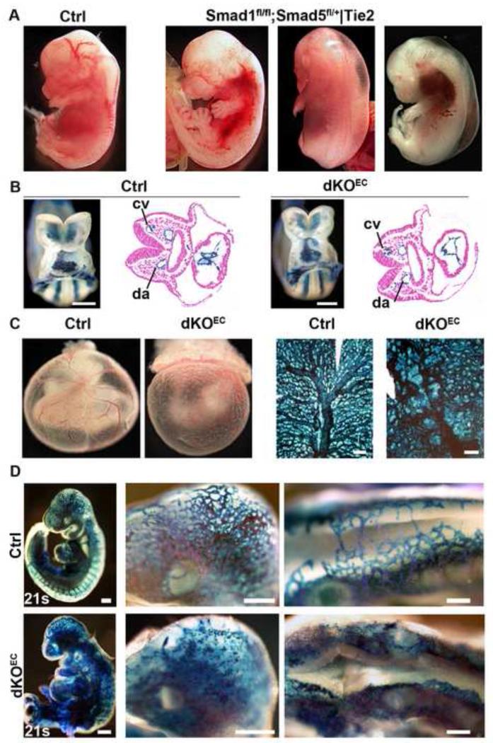
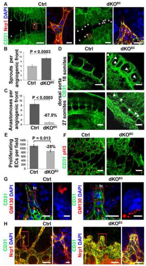
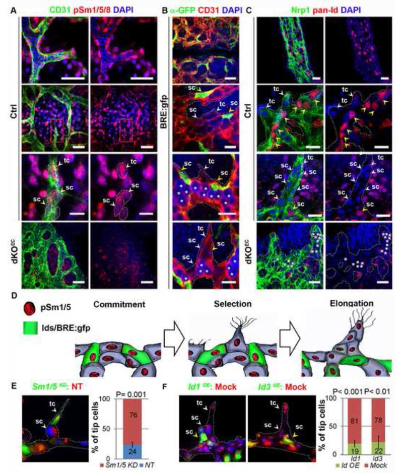
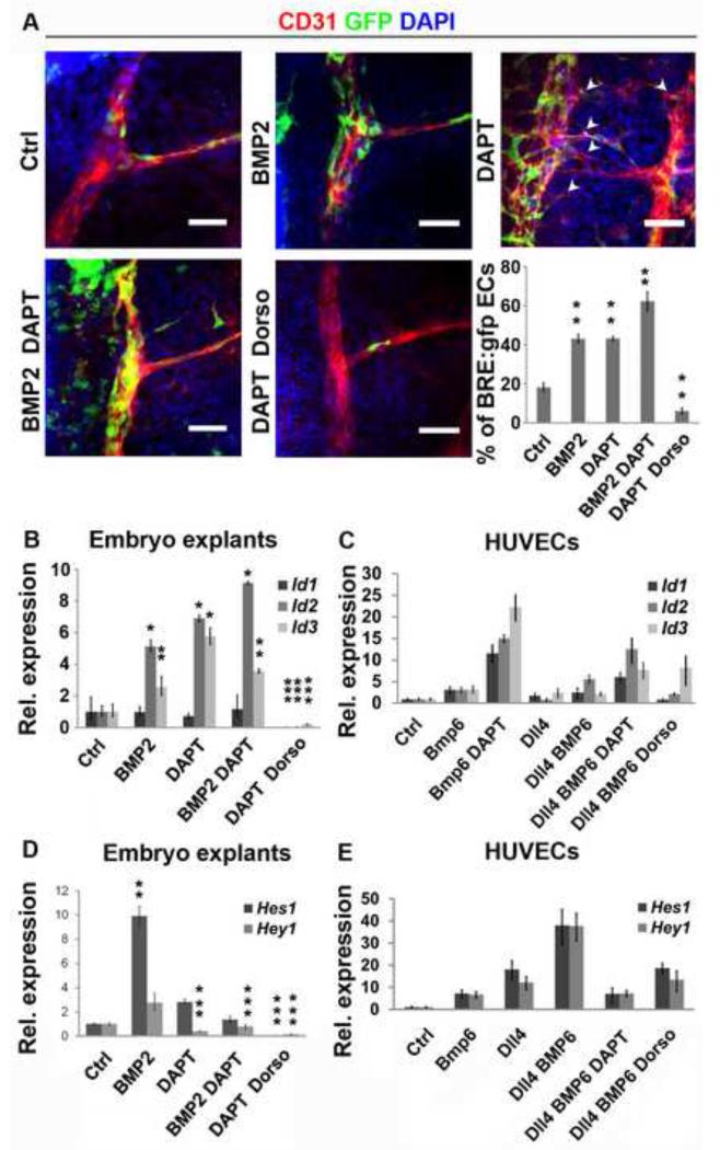
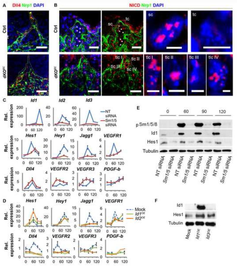
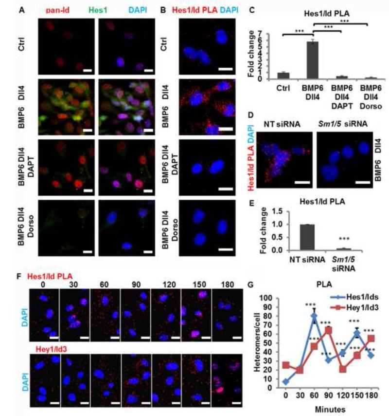
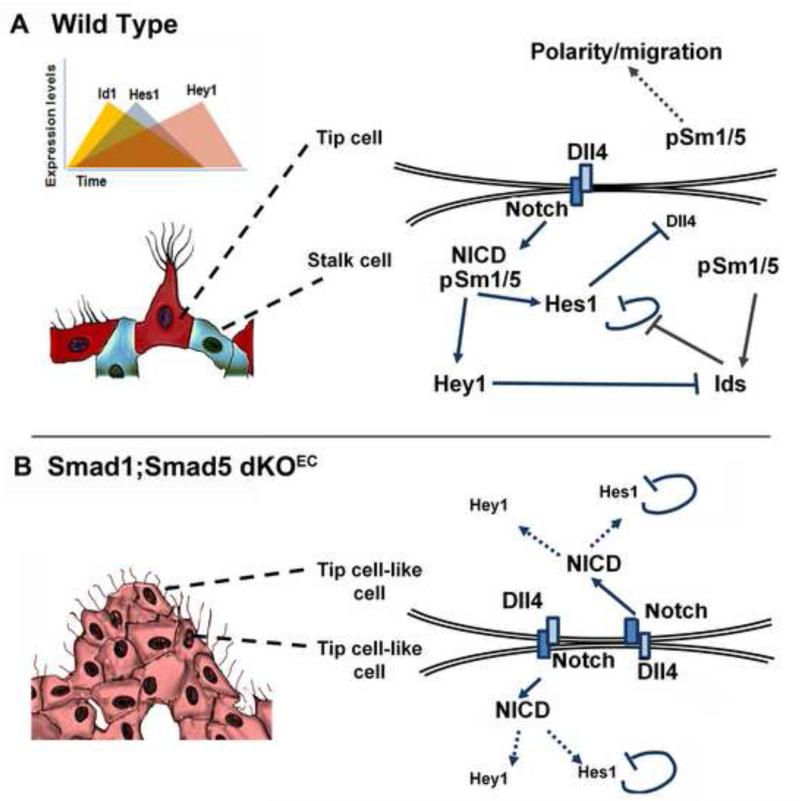
Gradients of vascular endothelial growth factor (VEGF) induce single endothelial cells to become leading tip cells of emerging angiogenic sprouts. Tip cells then suppress tip-cell features in adjacent stalk cells via Dll4/Notch-mediated lateral inhibition. We report here that Smad1/Smad5-mediated BMP signaling synergizes with Notch signaling during selection of tip and stalk cells. Endothelium-specific inactivation of Smad1/Smad5 in mouse embryos results in impaired Dll4/Notch signaling and increased numbers of tip-cell-like cells at the expense of stalk cells. Smad1/5 downregulation in cultured endothelial cells reduced the expression of several target genes of Notch and of other stalk-cell-enriched transcripts (Hes1, Hey1, Jagged1, VEGFR1, and Id1-3). Moreover, Id proteins act as competence factors for stalk cells and form complexes with Hes1, which augment Hes1 levels in the endothelium. Our findings provide in vivo evidence for a regulatory loop between BMP/TGFβ-Smad1/5 and Notch signaling that orchestrates tip- versus stalk-cell selection and vessel plasticity.


Figures

Figure 1
Figure 1. Normal vasculogenesis but impaired angiogenesis in dKOEC embryos
a, E13.5 control embryo (left panel) and embryos containing one functional allele of Smad1 or Smad5 in ECs. b-d, Embryos lacking the four alleles of Smad1 and Smad5 in endothelium. b, Control and mutant E8.5 whole-mount and sectioned R26R reporter embryo stained with X-gal (blue). c-d, Whole mount and flat mounted X-gal stained yolk sacs of control and mutant E9.5 R26R reporter embryos. In d, magnified views are shown of the head and roof of the hindbrain (middle and right panels). Abbreviations: cv, cardinal vein; da, dorsal aorta; s, somites. Scale bars: 250 μm (b,c,d left panels) and 300 μm (d middle and left panels). See also Figure S1.
Figure 2
Figure 2. Smad1/5 signaling regulates angiogenesis
a, Dorsal vascular plexus (anti-CD31/anti-Nrp1) in flat-mounted hindbrain roofs (E9.5). Asterisks and boxed areas (right panels) show sprouts quantified in (b). b,c, Quantification of sprouts (b) and dorsal anastomoses (c). d, Dorsal aortae (anti-CD31) from control and dKOEC E9.5 embryos, arrow heads show ectopic sprouts. e,f, Quantification of proliferation (phospho H3-positive ECs) in the dorsal vascular plexus. g, Position of the Golgi apparatus (anti-GM130) in tip cells. h, Filopodia (anti-Nrp1) in endothelium (anti-CD31). Boxed region in g-h are magnified in right panels. Abbreviations: tc, tip cell. Quantifications are averages from several embryos (b,c: n ≥ 21; e: n ≥ 11). Scale bars: 200 μm (a, left panels), 20 μm (a, boxed area; g, left panels); 150 μm (d,f,h, left panels); 4 μm (g, boxed area) and 50 μm (h, boxed area). See also Figure S2 and S3.
Figure 3
Figure 3. Smad1/5 signaling regulates stalk cell competence via Id proteins
a, Anti-p-Smad1/5/8 staining in endothelium of E9.5 control and dKOEC littermates. Nuclei of tip and stalk cells are highlighted by dashed lines. b, GFP localization (BRE:gfp) in early stalk cells (middle panels) and elongated sprouts (bottom panel), c, Id protein localization in wild-type yolk sac artery (upper panels) and embryonic vasculature (middle panels). Absence of Id proteins in mutant sprouts (bottom panels). Yellow and white arrowheads indicate pan-Id/GFP positive or negative cells respectively. Asterisks represent red blood cells. d, Schematic representation of the dynamic p-Sm1/5/8 and Id/BRE:gfp localization in developmental angiogenic sprouts. e, Chimaeric tube formation assay and quantification of Smad1/5KD (green, calcein) or WT (red, DiI-AC-LDL) leading “tip” cells. f, Chimaeric tube formation assay and quantification of Id1OE/Id3OE (green, calcein) or WT (red, DiI-AC-LDL) leading “tip” cells. Abbreviations: sc, stalk cell; tc, tip cell. Quantifications are averages from several sprout/tubular structures (e: n ≥ 50; f: n ≥ 49). Scale bars from top to bottom: 15 μm, 50 μm, 15 μm and 40 μm in (a); 50 μm, 15 μm, 15 μm and 15 μm in (b); 50 μm, 15 μm, 15 μm and 50 μm in (c). See also Figure S4.
Figure 4
Figure 4. Cross-signaling between Notch and Smad1/5 pathways
a, GFP localization in endothelium (anti-CD31) in E9.5 BRE:gfp dorsal explants exposed to different treatments. The arrows heads show ectopic intrasomitic vessels. Quantification of GFP positive ECs from dorsal aorta. b-e, mRNA expression analysis of Id1-3 in embryo explants (c) and HUVECs (d). Hes1 and Hey1 expression in embryo explants (c) and HUVECs (e). Scale bars: 50 μm. See also Figure S5.
Figure 5
Figure 5. Smad1/5 mediated regulation of Dll4/Notch signaling in endothelial cells
a,b Aberrant Dll4 (a) and NICD (b) localization in dKOEC angiogenic sprouts. In (b), left and middle panels are identical. Boxed areas of presumptive tip cells are magnified at the right. Asterisks represent red blood cells. c,d, Effect of Smad1/5 siRNA (c) or Id1/3 overexpression in the regulation of tip and stalk cell enriched transcripts in HUVECs. e,f, p-Smad1/5/8, Id1 and Hes1 proteins levels after Sm1/5 siRNA mediated downregulation (e) and Id1/3 overexpression (f) in HUVECs. Abbreviations: sc, stalk cell; tc, tip cell; tlc, tip cell-like cells. Scale bars from left to right: 25 μm in (a); 30 μm in (b) left and middle panels and 5 μm in right panels. See also Figures S6.
Figure 6
Figure 6. Interaction of Hes1 and Id proteins in ECs
a, Endogenous Hes1 and Id localization in HUVECs treated with Dll4, BMP6, DAPT or Dorsomorphin. b, Hes1/Ids heteromers detected by in situ PLA using pan-Id and Hes1 antibodies. c, PLA signal quantification of (b). d, e, Reduced levels of Hes1/Ids heteromers upon Sm1/5 siRNA transfection. f, g, Dynamic regulation of Hes1/Ids and Hey1/Id3 heteromers in function of time. P values were calculated using two-tailed Student’s t-test (c,d, n ≥ 71; g n, ≥ 105). Abbreviations: Dorso, dorsomorphin hydrochloride. Scale bars: 40 μm (a, f), 15 μm (b, d). See also Figures S7.
Figure 7
Figure 7. Schematic model of the role of Smad1 and Smad5 in Dll4/Notch mediated stalk cell behavior
a, In wild type embryos, the Dll4-rich leading tip cell (red) of the emerging sprout activates in adjacent cells higher levels of Notch signaling which directs them to acquire a stalk cell behavior (blue). Notch activation and target gene expression (Hey and Hes) downregulates the expression of VEGFR2, VEGFR3 and Dll4, and upregulates VEGFR1 resulting in stalk cell behavior. Downstream of Smad1/5 signaling, Id proteins release the negative autoregulatory loop of Hes1 in stalk cells. Conversely, increasing levels of the Notch target Hey1 will progressively inhibit Id steady-state levels, which will then indirectly cause attenuation of the Notch pathway by Hes1 downregulation. b, Absence of Id proteins in Smad1/5 deficient ECs will result in Hes1 downregulation. High levels of VEGFR2 and Dll4, and deficient Notch target gene expression in stalk cells turn them into a tip cell-like cell phenotype (pink) and prevents the downregulation of Notch1 signaling (nuclear NICD accumulation) in the leading ‘tip’ cells. The failure to balance tip versus stalk cell ratios and to transiently stabilize stalk cells, together with impaired tip cell polarity and directed migration, is likely to cause excessive sprouting and vessel coalescence in mutant embryos.

References

    1. Bai G, Sheng N, Xie Z, Bian W, Yokota Y, Benezra R, Kageyama R, Guillemot F, Jing N. Id sustains Hes1 expression to inhibit precocious neurogenesis by releasing negative autoregulation of Hes1. Dev Cell. 2007;13:283–297. - PubMed
    1. Bentley K, Mariggi G, Gerhardt H, Bates PA. Tipping the balance: robustness of tip cell selection, migration and fusion in angiogenesis. PLoS Comput Biol. 2009;5:e1000549. - PMC - PubMed
    1. Chang H, Huylebroeck D, Verschueren K, Guo Q, Matzuk MM, Zwijsen A. Smad5 knockout mice die at mid-gestation due to multiple embryonic and extraembryonic defects. Development. 1999;126:1631–1642. - PubMed
    1. Dahlqvist C, Blokzijl A, Chapman G, Falk A, Dannaeus K, Ibanez CF, Lendahl U. Functional Notch signaling is required for BMP4-induced inhibition of myogenic differentiation. Development. 2003;130:6089–6099. - PubMed
    1. David L, Feige JJ, Bailly S. Emerging role of bone morphogenetic proteins in angiogenesis. Cytokine Growth Factor Rev. 2009;20:203–212. - PubMed

Publication types

MeSH terms

Substances

LinkOut - more resources