. 2011 Oct-Dec;26(4):229-48.

Screening for chronic cerebrospinal venous insufficiency (CCSVI) using ultrasound: recommendations for a protocol

Affiliations

Screening for chronic cerebrospinal venous insufficiency (CCSVI) using ultrasound: recommendations for a protocol

A N Nicolaides et al. Funct Neurol. 2011 Oct-Dec.

Abstract

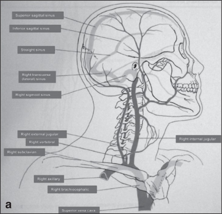
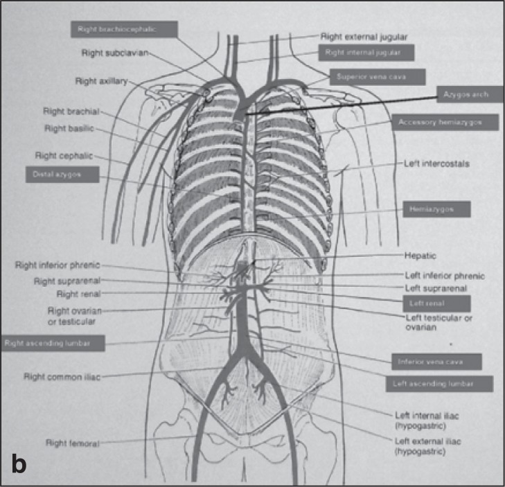
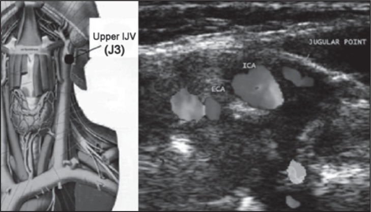
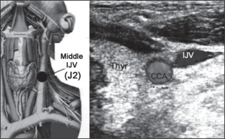
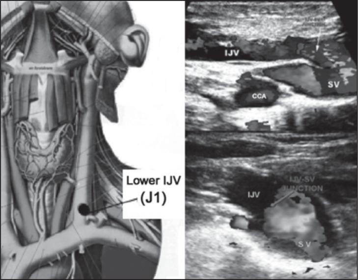
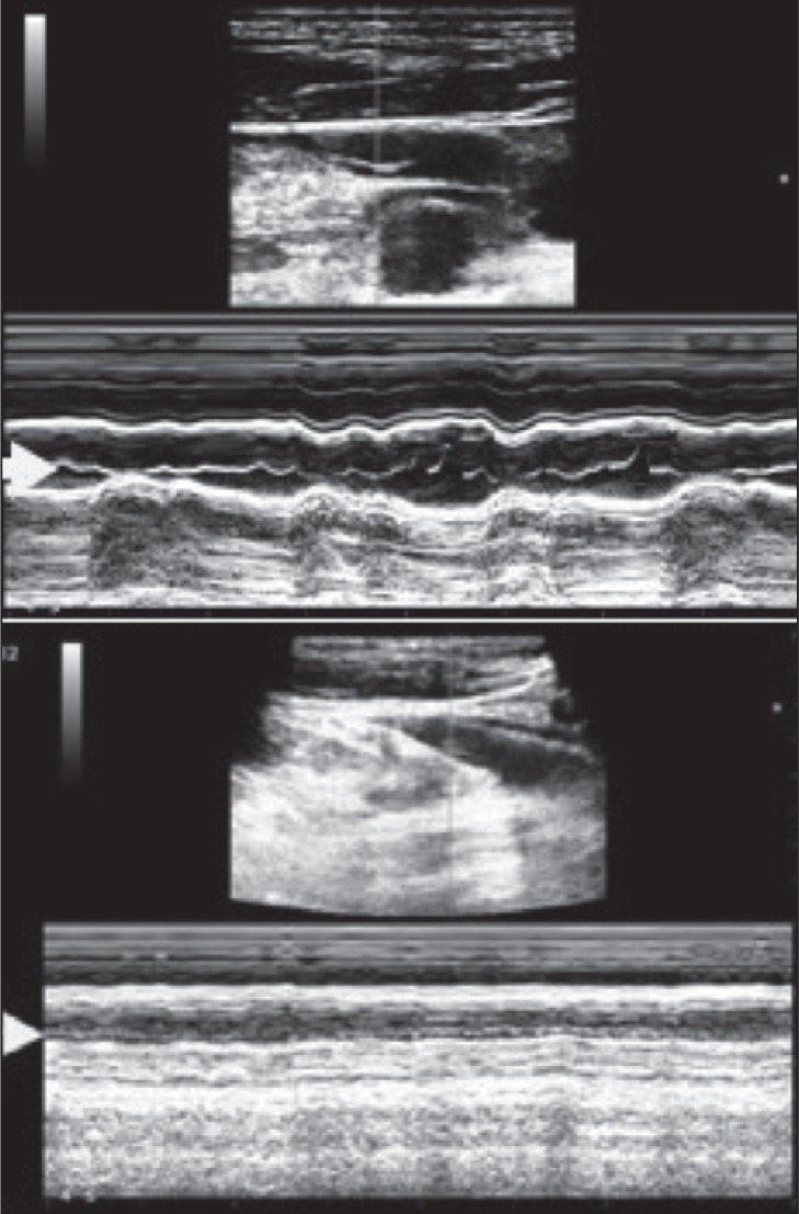
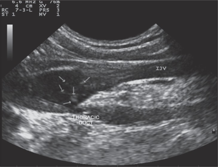
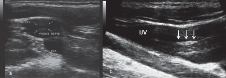
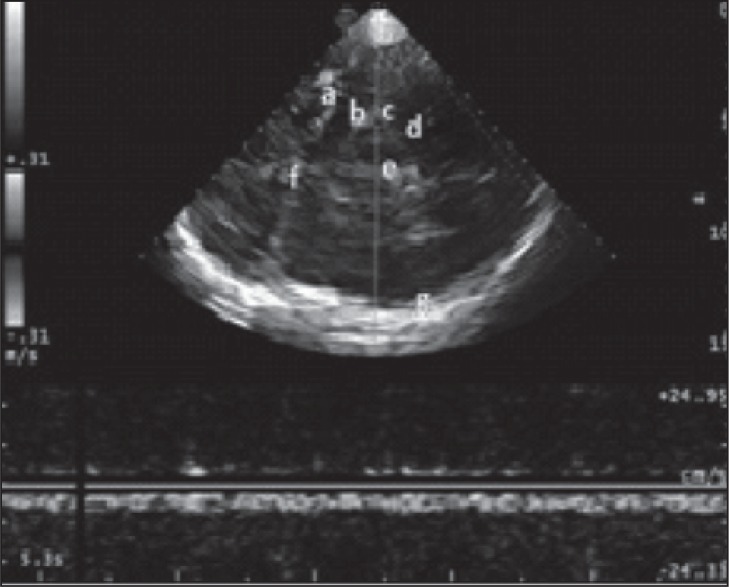
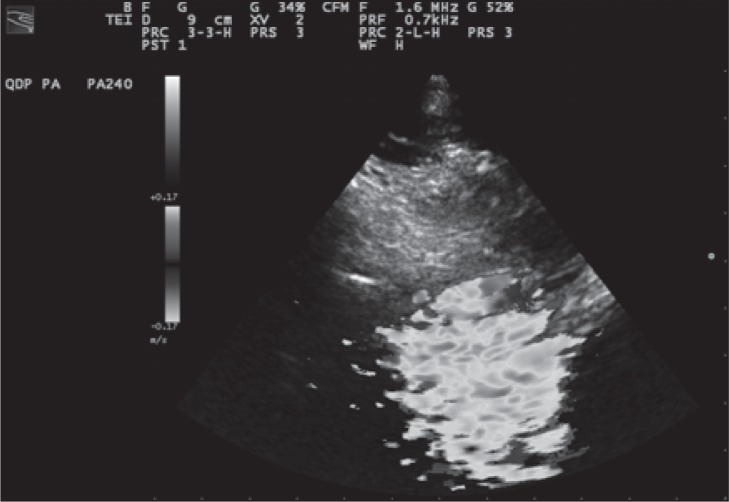
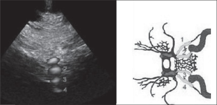
Chronic cerebrospinal venous insufficiency (CCSVI) is a syndrome characterized by stenoses or obstructions of the internal jugular and/or azygos veins with disturbed flow and formation of collateral venous channels. Ultrasound and venographic studies of the internal jugular and azygos venous systems in patients with multiple sclerosis (MS) have demonstrated a high prevalence of CCSVI (mean 71%, range 0-100%; n=1336) associated with activation of collaterals. By contrast, ultrasound and venographic examinations of normal controls and patients without MS have demonstrated a much lower prevalence (mean 7.1%, range 0-22%; n=505). Ultrasound in the form of duplex scanning uses a combination of physiological measurements as well as anatomical imaging and has been used for the detection of CCSVI by different centers with variable results. A high prevalence of obstructive lesions, ranging from 62% to 100%, has been found by some teams in patients with MS compared with a low prevalence (0-25%) in controls. However, others have reported absence of these lesions or a lower prevalence (16-52%). This variability could be the result of differences in technique, training, experience or criteria used. In order to ensure a high reproducibility of duplex scanning with comparable accuracy between centers a detailed protocol with standard methodology and criteria is needed. Also, standardization of the method of reporting of duplex measurements and other findings will facilitate validation of the proposed criteria by different centers. The aim of this document is to produce recommendations for such a protocol and indicate what future research is needed in order to address areas of uncertainty.


Figures

Figures 1a and b
Figures 1a and b
The cerebrospinal veins are a highly complex system rich in anastomoses, extending from the intracranial and intravertebral veins to the neck and the chest, with collaterals also in the retroperitoneal veins.
Figures 1a and b
Figures 1a and b
The cerebrospinal veins are a highly complex system rich in anastomoses, extending from the intracranial and intravertebral veins to the neck and the chest, with collaterals also in the retroperitoneal veins.
Figure 2
Figure 2
Left: Upper IJV level (J3) in an anatomical plate modified from the Sobotta Atlas of Anatomy. Right: transverse scan at the upper (J3) level; ECA=external carotid artery; ICA=internal carotid artery. The jugular point corresponds to the position of the internal jugular vein.
Figure 3
Figure 3
Left: Middle IJV level (J2) in an anatomical plate modified from the Sobotta Atlas of Anatomy. Right: transverse scan at the middle (J2) level; CCA=common carotid artery; IJV=internal jugular vein; Thyr=thyroid gland.
Figure 4
Figure 4
Left: Lower IJV level (J1) in an anatomical plate modified from the Sobotta Atlas of Anatomy. Top right: longitudinal scan at the lower (J1) level; IJV=internal jugular vein; SV=subclavian vein; CCA=common carotid artery. Bottom right: image of the junction in the transverse plane. In both cases, the arrows indicate the junction.
Figure 5
Figure 5
Top: M-mode evaluation of the jugular valve showing mobility of the leaflets as indicated by the arrow. Bottom: immobility of valve leaflets demonstrated in M-mode (arrow).
Figure 6
Figure 6
B-mode anomaly artifact created by the presence of streams of lymph within the venous blood flow of the IJV.
Figure 7a
Figure 7a
B-mode artifact created by the vagus nerve, in the transverse view at the lower IJV level (J1). The arrows indicate the hyperechoic thickening of the neural guaina and fibers. Figure 7b - Mirror artifact, in the longitudinal aspect of the IJV.
Figure 8
Figure 8
Bidirectional flow in the IJV seen in the transverse view needs to be confirmed by longitudinal scans and Doppler waveform recordings ( Fig.s 10–12 ).
Figure 9
Figure 9
Bidirectional flow in the IJV seen in longitudinal view. Red color indicates reflux.
Figure 10
Figure 10
Bidirectional Doppler velocities in the IJV recorded with a large sample volume.
Figure 11
Figure 11
A longitudinal image showing a long-lasting reflux. Multi-angle Doppler makes it possible to record the direction of flow in the internal jugular vein (IJV) and in the common carotid artery (CCA) simultaneously.
Figure 12
Figure 12
Example of increased flow velocity up to 119 cm/sec in the IJV, and absence of reflux. This hemodynamic pattern is not a CCSVI criterion per se, but is highly suggestive of stenosis.
Figure 13
Figure 13
Example of absence of detectable flow in longitudinal scan of an IJV in supine position, and PRF is 1.2 kHz.
Figure 14
Figure 14
Blood from the IJV is shunted into the thyroid vein, a collateral circulation activated by the presence of obstruction at the lower IJV (J1). Note the enlarged veins in the thyroid gland. CCA=common carotid artery; C COLL= collateral circle.
Figure 15
Figure 15
Top: Example of cross-sectional area (CSA) measurement of the IJV in supine position. Bottom: Example of CSA measurement of the IJV in sitting position and relative calculation of ΔCSA. In healthy individuals the CSA in the supine position is greater than in the upright position. CSGS=cross sectional jugular area in sitting position; CCA=common carotid artery.
Figure 16
Figure 16
Example of color Doppler vertebral vein assessment. CCA=common carotid artery; VV=vertebral vein; VA=vertebral artery.
Figure 17
Figure 17
Example of a “blocked” vertebral vein (VV), confirmed by Doppler spectrum analysis (indicated by absence of flow at the arrow). VV=vertebral vein; VA=vertebral artery.
Figure 18a
Figure 18a
Example of reflux: the blood flow at the level of the superior petrosal sinus (SPS) shows opposite directions between inspiration and expiration; this finding shows the presence of reversed blood flow within the examined vessel between the two phases of respiration. The blood flow direction of the inferior petrosal sinus (IPS) is not detectable during expiration. The blood flow direction of the contralateral inferior petrosal sinus (CIPS) is not detectable during inspiration or expiration.
Figure 18b
Figure 18b
Example of no reflux: the blood flow at the level of the superior petrosal sinus (SPS) shows the same direction between inspiration and expiration; the blood flow direction of the inferior petrosal sinus (IPS) is not detectable during expiration; the blood flow direction of the contralateral inferior petrosal sinus (CIPS) is not detectable during inspiration or expiration.
Figure 1
Figure 1
Transtemporal approach at the level of midbrain (e) contralateral skull (g). The ultrasonic beam makes it possible to identify: the middle cerebral artery (a) and the contralateral skull (f), the posterior cerebral artery, pre-peduncular part (b), the Rosenthal vein (c) and the posterior cerebral artery, post-peduncular part (d).
Figure 2
Figure 2
Color cloud artifact seen from the transcranial window at the level of the condyloid process of the mandible (PRF 0.7 kHz). This artifact represents a really powerful “Doppler” anatomical marker in order to find the plane in which one or more of the cerebral veins we want to examine are positioned.
Figure 3
Figure 3
Visualization of the power Doppler signal (instead of color Doppler for increased spatial resolution) of the superior petrosal sinus ( 1 ), inferior petrosal sinus ( 2 ), contralateral inferior petrosal sinus ( 3 ) and (partially) contralateral superior petrosal sinus ( 4 ), from the transcranial window at the level of the condyloid process of the mandible.

References

    1. Zamboni P , Galeotti R , Menegatti E , et al. Chronic cerebrospinal venous insufficiency in patients with multiple sclerosis . J Neurol Neurosurg Psychiatry . 2009 ; 80 : 392 – 399 . - PMC - PubMed
    1. Zamboni P , Consorti G , Galeotti R , et al. Venous collateral circulation of the extracranial cerebrospinal outflow routes . Curr Neurovasc Res . 2009 ; 6 : 204 – 212 . - PubMed
    1. Zivadinov R , Galeotti R , Hojnacki D , et al. Value of MR venography for detection of internal jugular vein anomalies in multiple sclerosis: a pilot longitudinal study . AJNR Am J Neuroradiol . 2011 ; 32 : 938 – 946 . - PMC - PubMed
    1. Zivadinov R , Marr K , Cutter G , et al. Prevalence, sensitivity, and specificity of chronic cerebrospinal venous insufficiency in MS . Neurology . 2011 ; 77 : 138 – 144 . - PubMed
    1. Al-Omari MH , Rousan LA . Internal jugular vein morphology and hemodynamics in patients with multiple sclerosis . Int Angiol . 2010 ; 29 : 115 – 120 . - PubMed

Publication types

MeSH terms

LinkOut - more resources