

Increased expression of macrophage colony-stimulating factor and its receptor in patients with endometriosis
- PMID: 22365076
- PMCID: PMC3583230
- DOI: 10.1016/j.fertnstert.2012.02.007
Increased expression of macrophage colony-stimulating factor and its receptor in patients with endometriosis
Abstract
Objective: To investigate the expression and regulation of colony-stimulating factor 1 (CSF-1) and its receptor, C-FMS, in endometriosis.
Design: In vivo and vitro study.
Setting: University-based academic medical center.
Patient(s): Reproductive-age women undergoing surgery for benign conditions.
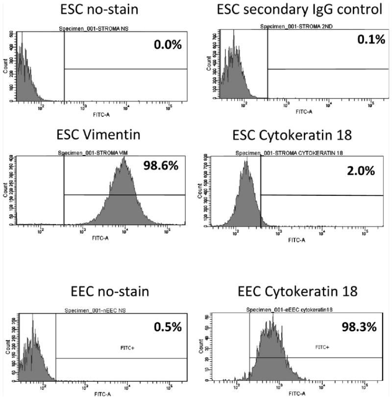
Intervention(s): Peritoneal and endometrial tissue samples were obtained.
Main outcome measure(s): CSF-1 and C-FMS expression.
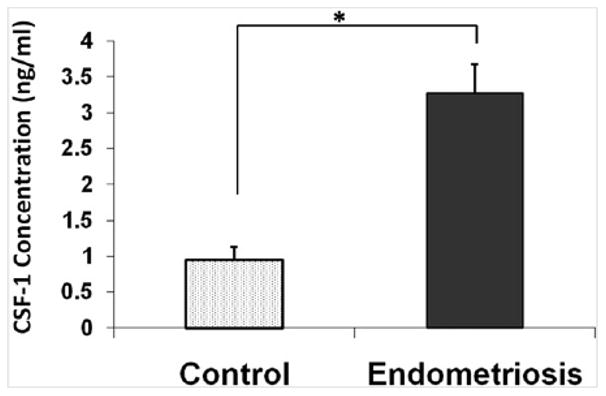
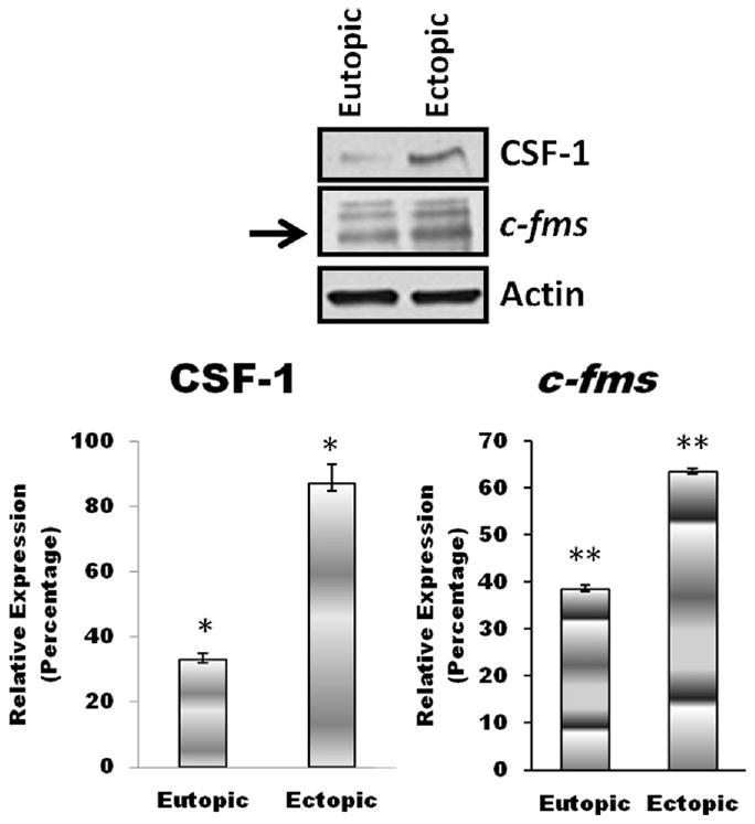
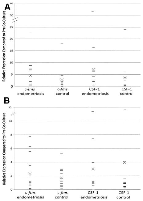
Result(s): Significantly higher CSF-1 levels were found in peritoneal fluid of patients with endometriosis compared with control subjects. Ectopic endometriotic tissue had 3.5-fold and 1.7-fold increases in CSF-1 and C-FMS expression, respectively, compared with eutopic tissue. Coculture of endometrial cells from either established cell lines or patient samples with peritoneal mesothelial cells (PMCs) led to increased expression of CSF-1 and C-FMS. A higher but nonsignificant increase in levels of C-FMS and CSF-1 was found in cocultures of endometrial epithelial cells from patients with endometriosis compared with those without endometriosis.
Conclusion(s): Increased CSF-1 levels may contribute to endometriosis lesion formation and progression. Elevation in CSF-1 after coculture of endometrial cells with PMCs suggests that endometrial tissue may be a source of peritoneal CSF-1. Increased C-FMS expression in endometrial cells from women with endometriosis cocultured with PMCs suggests that endometrial tissue involved in lesion formation is highly responsive to CSF-1 signaling.
Copyright © 2012 American Society for Reproductive Medicine. Published by Elsevier Inc. All rights reserved.
Figures
References
-
- Eskenazi B, Warner ML. Epidemiology of endometriosis. Obstet Gynecol Clin North Am. 1997;24:235–58. - PubMed
-
- Olive DL, Henderson DY. Endometriosis and mullerian anomalies. Obstet Gynecol. 1987;69:412–5. - PubMed
-
- D′Hooghe TM, Debrock S. Endometriosis, retrograde menstruation and peritoneal inflammation in women and in baboons. Hum Reprod Update. 2002;8:84–8. - PubMed
-
- Halme J, Hammond MG, Hulka JF, Raj SG, Talbert LM. Retrograde menstruation in healthy women and in patients with endometriosis. Obstet Gynecol. 1984;64:151–4. - PubMed
Publication types
MeSH terms
Substances
Grants and funding
LinkOut - more resources
Full Text Sources
Medical
Research Materials
Miscellaneous
