. 2012 Apr;245(1-2):1-7.
doi: 10.1016/j.jneuroim.2012.01.005. Epub 2012 Feb 25.

Interleukin-10 plays a crucial role in suppression of experimental autoimmune encephalomyelitis by Bowman-Birk inhibitor

Affiliations

Interleukin-10 plays a crucial role in suppression of experimental autoimmune encephalomyelitis by Bowman-Birk inhibitor

Hong Dai et al. J Neuroimmunol. 2012 Apr.

Abstract

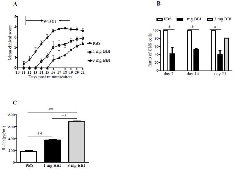
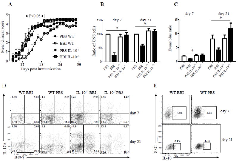
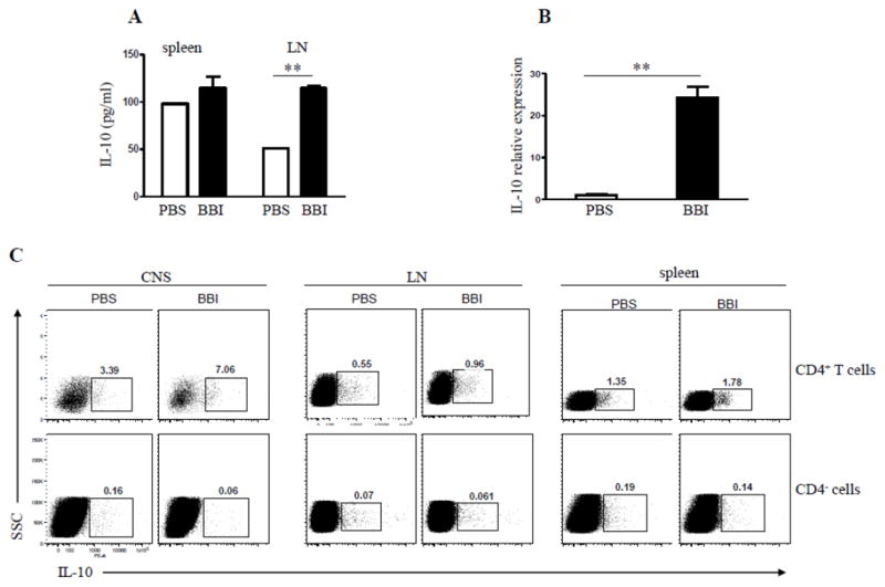
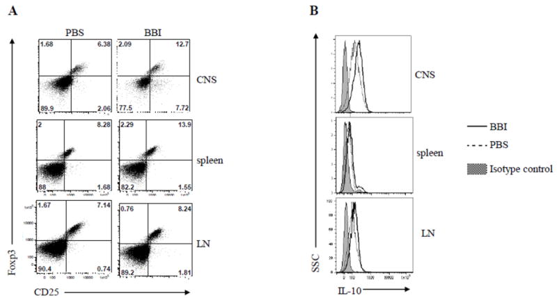
The Bowman-Birk inhibitor (BBI) is a soybean-derived serine protease inhibitor with anti-inflammatory properties. Experimental autoimmune encephalomyelitis (EAE) serves as an animal model of the central nervous system (CNS) inflammatory disorder multiple sclerosis (MS). EAE is mediated by Th1 and Th17 cells which migrate into the CNS and initiate inflammation directed against myelin components, resulting in CNS pathology and neurological clinical deficit. We have shown previously that oral treatment with BBI delays onset of EAE and reduces its severity. These beneficial effects were associated with an increase in IL-10 secretion by immune cells of BBI-treated mice. It is not known, however, whether this was a causal relationship or simply an epiphenomenon. In the present study we provide evidence that BBI regulates CD4+ T cell immune responses in EAE. BBI administration delayed the onset of EAE and reduced its severity in an IL-10-dependent manner, as BBI-mediated suppression of EAE was abrogated in IL-10 knockout mice. The beneficial effects were accompanied by reduced IFN-γ, IL-17 and increased IL-10 production, as well as increased Foxp3 expression. CD4+ T cells were the major source of IL-10 in the periphery and in the CNS during BBI treatment. Furthermore, BBI-treated mice had reduced numbers of infiltrated cells in the CNS, including Th17 cells, as compared with PBS-treated control animals. In conclusion, our data provide clear evidence for the essential role of IL-10 in BBI-mediated suppression in EAE, and indicate that BBI may be a promising candidate for the development of a novel MS therapy.


Figures

Figure 1
Figure 1. BBI suppresses EAE in C57BL/6 mice
(A) C57BL/6 mice were immunized with MOG35-55 and treated with BBI (1 mg/day, n=10 or 3 mg/day, n=4) or PBS (n=11) by oral gavage from the day of immunization until the end of the experiment; mice were scored daily for clinical disease. (B) On days 7, 14 and 21 p.i., mice were sacrificed and cells from CNS isolated and counted. Data are shown as percentage relative to the cell number obtained from PBS-treated mice. (C) On day 21 p.i., draining LN cells were harvested and stimulated with anti-CD3/CD28 for 3 days; thereafter supernatants were analyzed for IL-10 by ELISA. P values refer to the comparison between BBI- vs. PBS-treated groups. Data are representative of three independent experiments.
Figure 2
Figure 2. BBI-mediated suppression of EAE is abated in IL-10-/- mice
(A) WT and IL-10-/- mice on C57BL/6 background were immunized with MOG35-55 and treated with 1 mg/day of BBI or PBS (n=12 in each group) by oral gavage from the day of immunization until the end of the experiment. Mice were scored daily for clinical disease. (B) WT and IL-10-/- mice (n=12 in each group) were immunized with MOG35-55 and treated with BBI (1 mg/day) or PBS by oral gavage from the day of immunization. Mice were sacrificed on days 7 and 21 p.i.; cells from CNS were isolated and counted. Data are shown as a percentage relative to the cell number obtained from WT PBS-treated mice. (C) The integrity of BBB was evaluated by measurement of Evans blue content in brains at days 7 and day 20 p.i. (n=6 per group). Results are presented as a ratio between background levels obtained from naïve mice injected with Evans blue and mice with EAE. Values represent the mean ± S.E.M. (D) Mononuclear cells were isolated from spinal cords on days 7 and 21 p.i., stained and analyzed by flow cytometry for IL-17A and IFN-γ production. CD4+ T cells are shown. (E) Mononuclear cells were isolated from spinal cords on days 7 and 21 p.i., stained and analyzed by flow cytometry for IL-10 production. CD4+ T cells are shown. P value refers to comparison between BBI-treated WT mice and other groups. Data are representative of two independent experiments.
Figure 3
Figure 3. CD4+ T cells are the main source of IL-10 following BBI treatment
C57BL/6 mice were immunized with MOG35-55 and treated daily with BBI (1 mg/day) or PBS by oral gavage from day 0 p.i. and sacrificed on day 7 p.i. (A) LN cells were stimulated with MOG35-55 for 3 days, and concentrations of IL-10 measured in the supernatants by ELISA. (B) C57BL/6 mice (n=6 per group) were immunized with MOG35-55 and treated daily with BBI (1 mg/day) or PBS by oral gavage from the day of immunization. Mice were sacrificed on day 14 p.i.; mononuclear cells were isolated from the CNS and used to isolate mRNA. mRNA levels of IL-10 were determined by quantitative real-time PCR. Data are representative of three independent experiments. (C) Spinal cord, splenic and LN cells of immunized mice that had been treated for 14 days p.i. with BBI were isolated and analyzed for intracellular production of IL-10 by flow cytometry. ** p < 0.01. Data are representative of three independent experiments.
Figure 4
Figure 4. BBI treatment induces CD4+CD25+ Foxp3+ Tregs
C57BL/6 mice (n=6 per group) were immunized with MOG35-55 and treated with BBI (1mg/day) or PBS by oral gavage from day 0 p.i.; mice were sacrificed on day 14 p.i. (A) CD4+ T cells from spinal cord, spleen and LN were gated and their CD25 and Foxp3 expression analyzed by flow cytometry. (B) Flow cytometric analysis of IL-10 intracellular levels in CD4+CD25+ T cells from spinal cord, spleen and LN of mice treated with BBI for 14 days p.i. Data are representative of three experiments.

Similar articles

Cited by

References

    1. Akirav EM, Bergman CM, Hill M, Ruddle NH. Depletion of CD4(+)CD25(+) T cells exacerbates experimental autoimmune encephalomyelitis induced by mouse, but not rat, antigens. J Neurosci Res. 2009;87:3511–3519. - PMC - PubMed
    1. Anderson AC, Reddy J, Nazareno R, Sobel RA, Nicholson LB, Kuchroo VK. IL-10 plays an important role in the homeostatic regulation of the autoreactive repertoire in naive mice. J Immunol. 2004;173:828–834. - PubMed
    1. Anderton SM. Treg and T-effector cells in autoimmune CNS inflammation: a delicate balance, easily disturbed. Eur J Immunol. 2010;40:3321–3324. - PubMed
    1. Axtell RC, de Jong BA, Boniface K, van der Voort LF, Bhat R, De Sarno P, Naves R, Han M, Zhong F, Castellanos JG, Mair R, Christakos A, Kolkowitz I, Katz L, Killestein J, Polman CH, de Waal Malefyt R, Steinman L, Raman C. T helper type 1 and 17 cells determine efficacy of interferon-beta in multiple sclerosis and experimental encephalomyelitis. Nat Med. 2010;16:406–412. - PMC - PubMed
    1. Bettelli E, Das MP, Howard ED, Weiner HL, Sobel RA, Kuchroo VK. IL-10 is critical in the regulation of autoimmune encephalomyelitis as demonstrated by studies of IL-10- and IL-4-deficient and transgenic mice. J Immunol. 1998;161:3299–3306. - PubMed

Publication types

MeSH terms