. 2012 Mar;95(3):1188-97.
doi: 10.3168/jds.2011-4430.

Characterization of bovine glucose transporter 1 kinetics and substrate specificities in Xenopus oocytes

Affiliations

Characterization of bovine glucose transporter 1 kinetics and substrate specificities in Xenopus oocytes

P A Bentley et al. J Dairy Sci. 2012 Mar.

Abstract

Glucose is an essential substrate for lactose synthesis and an important energy source in milk production. Glucose uptake in the mammary gland, therefore, plays a critical role in milk synthesis. Facilitative glucose transporters (GLUT) mediate glucose uptake in the mammary gland. Glucose transporter 1 (GLUT1) is the major facilitative glucose transporter expressed in the bovine mammary gland and has been shown to localize to the basolateral membrane of mammary epithelial cells. Glucose transporter 1 is, therefore, thought to play a major role in glucose uptake during lactation. The objective of this study was to determine the transport kinetic properties and substrate specificity of bovine GLUT1 using the Xenopus oocyte model. Bovine GLUT1 (bGLUT1) was expressed in Xenopus oocytes by microinjection of in vitro transcribed cRNA and was found to be localized to the plasma membrane, which resulted in increased glucose uptake. This bGLUT1-mediated glucose uptake was dramatically inhibited by specific facilitative glucose transport inhibitors, cytochalasin B, and phloretin. Kinetic analysis of bovine and human GLUT1 was conducted under zero-trans conditions using radio-labeled 2-deoxy-D-glucose and the principles of Michaelis-Menten kinetics. Bovine GLUT1 exhibited a Michaelis constant (K(m)) of 9.8 ± 3.0mM for 2-deoxy-d-glucose, similar to 11.7 ± 3.7 mM for human GLUT1. Transport by bGLUT1 was inhibited by mannose and galactose, but not fructose, indicating that bGLUT1 may also be able to transport mannose and galactose. Our data provides functional insight into the transport properties of bGLUT1 in taking up glucose across mammary epithelial cells for milk synthesis.


Figures

Figure 1
Figure 1
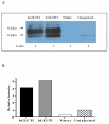
Expression of GLUT1 protein in Xenopus oocytes. Western blot analysis (A) was performed on homogenate of oocytes injected with either human GLUT1 (hGLUT1) or bovine GLUT1 (bGLUT1) cRNA. Water injected and uninjected oocytes were used for detection of endogenous GLUT1 expression. 75 μg of total protein were loaded in each lane. Relative GLUT1 band intensities were determined by densitometry (B).
Figure 2
Figure 2
Immunofluorescence staining of exogenous GLUT1 in Xenopus oocytes injected with either bovine GLUT1 cRNA (A) or human GLUT1 cRNA (B). Scale bar = 50 μm.
Figure 3
Figure 3
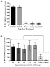
Transport activity of exogenous GLUT1 in Xenopus oocytes. A: Xenopus oocytes were injected with human GLUT1 (hGLUT1) or bovine GLUT1 (bGLUT1) cRNA (15 ng), water or were not injected (Uninj.). 100μM cytochalasin B (CCB) was added to the uptake reaction of a group of uninjected oocytes (Uninj. + CCB). Oocytes were exposed to 5 mM 2-DG containing 1 μCi/mL [3H]-2-DG for 15 minutes. Means of 2-DG uptake in the cRNA-injected oocytes versus water-injected or uninjected controls were compared using Tukey-Kraner HSD test, error bars represent SEM, p < 0.001. B: Oocytes were injected with 0.0015-45 ng of bovine GLUT1 cRNA and subjected to the transport analysis as in A. Data represent the uptake in the cRNA-injected oocytes after subtracting the uptake in water-injected oocytes. Statistical significance was determined by Welch ANOVA, p < 0.001 (***), comparing groups as indicated in the Figure.
Figure 4
Figure 4
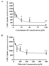
Inhibition of bovine GLUT1-mediated 2-deoxy-D-glucose uptake by cytochalsin B and phloretin in Xenopus oocytes. Oocytes were injected with bovine GLUT1 cRNA or water. Oocyte 2-DG uptake was measured after exposure of oocytes to 5 mM 2-DG and 1 μCi/mL [3H]-2-DG for 15 minutes and various concentrations of cytochalasin B (A) or phloretin (B). 2-DG uptake from water-injected oocytes was subtracted from cRNA injected oocytes. One phase non-linear decay model was used to identify the inhibition trend. Statistical analysis was conducted using Dunnett’s ANOVA, with 0 μM cytochalasin B or phloretin as the control group. Statistical significance was observed at p < 0.05 (*) and p < 0.01(**), p < 0.001 (***).
Figure 5
Figure 5
Effect of sodium pyruvate as an energy source on 2-deoxy-D-glucose uptake in Xenopus oocytes. Oocytes were injected with bovine or human GLUT1 cRNA or water. All oocytes were incubated for 72 hours in the presence (black bars) or absence (dashed bars) of 2.5 mM sodium pyruvate in Barth’s media. Oocytes were exposed to 5 mM 2-DG and 1 μCi/mL [3H]-2-DG for 15 minutes. 2-DG uptake in water-injected oocytes was subtracted from cRNA injected oocytes. Statistical analysis was conducted using the Tukey-Kramer HSD test; error bars represent SEM.
Figure 6
Figure 6
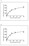
Kinetic analysis of 2-deoxy-D-glucose uptake by bovine (A) and human GLUT1 (B) in Xenopus oocytes. Oocytes were injected with bovine or human GLUT1 cRNA or water and exposed to various 2-DG concentrations containing 3 μCi/mL [3H]-2-DG for 15 minutes. Points represent 2-DG uptake in GLUT1-injected oocytes after correction for the uptake into water-injected oocytes. One representative assay is shown for each bGLUT1 and hGLUT1. Michaelis-Menten non-linear analysis was conducted in GraphPad Prism 5. Error bars represent SEM.
Figure 7
Figure 7
Inhibition of bovine GLUT1-mediated uptake of 2-deoxy-D-glucose by hexose substrates in Xenopus oocytes. Oocytes were injected with bovine GLUT1 cRNA or water and were exposed to 30 mM of each inhibitor sugar and 5 mM 2-DG and 1 μCi/mL [3H]-2-DG for 15 minutes. 2-DG uptake from water-injected oocytes was subtracted from cRNA-injected oocytes. Statistical analysis was conducted using Dunnett’s ANOVA, with L-glucose as the control group. Statistical significance was observed at p < 0.01(**), p < 0.001 (***), error bars represent SEM.

References

    1. Ahmed N, Berridge MV. N-glycosylation of glucose transporter-1 (Glut-1) is associated with increased transporter affinity for glucose in human leukemic cells. Leuk. Res. 1999;23:395–401. - PubMed
    1. Asano T, Katagiri H, Takata K, Lin JL, Ishihara H, Inukai K, Tsukuda K, Kikuchi M, Hirano H, Yazaki Y, et al. The role of N-glycosylation of GLUT1 for glucose transport activity. J. Biol. Chem. 1991;266:24632–24636. - PubMed
    1. Burant CF, Bell GI. Mammalian facilitative glucose transporters: evidence for similar substrate recognition sites in functionally monomeric proteins. Biochemistry. 1992;31:10414–10420. - PubMed
    1. Colville CA, Seatter MJ, Jess TJ, Gould GW, Thomas HM. Kinetic analysis of the liver-type (GLUT2) and brain-type (GLUT3) glucose transporters in Xenopus oocytes: substrate specificities and effects of transport inhibitors. Biochem. J. 1993;290:701–706. - PMC - PubMed
    1. Corpe CP, Bovelander FJ, Munoz CM, Hoekstra JH, Simpson IA, Kwon O, Levine M, Burant CF. Cloning and functional characterization of the mouse fructose transporter, GLUT5. Biochim. Biophys. Acta. 2002;1576:191–197. - PubMed

Publication types

LinkOut - more resources