. 2012 Feb 23;73(4):685-97.
doi: 10.1016/j.neuron.2011.11.033.

Propagation of tau pathology in a model of early Alzheimer's disease

Affiliations

Propagation of tau pathology in a model of early Alzheimer's disease

Alix de Calignon et al. Neuron. .

Erratum in

  • Neuron. 2012 Oct 18;76(2):461

Abstract

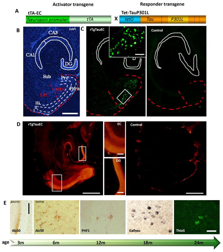
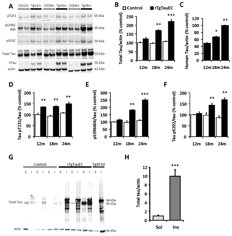
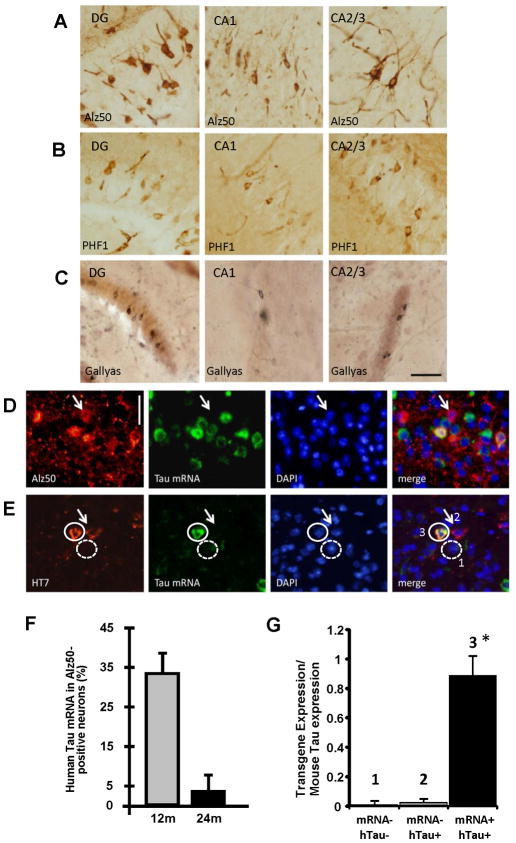
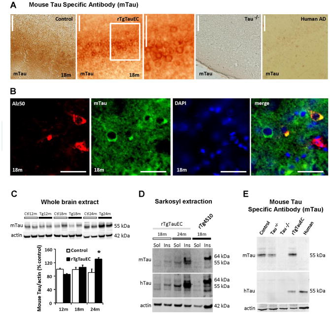
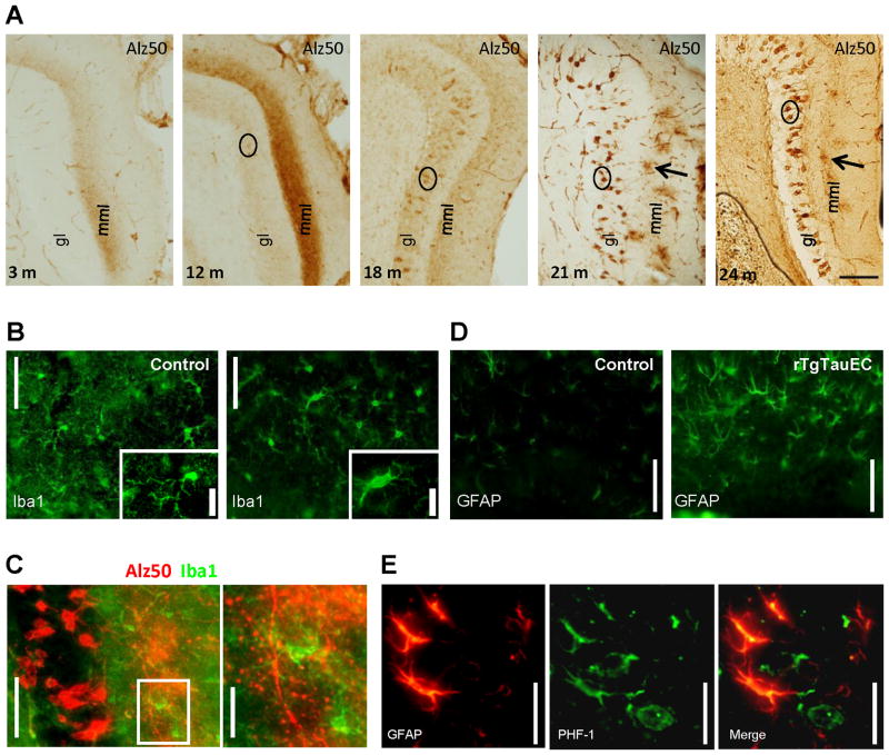
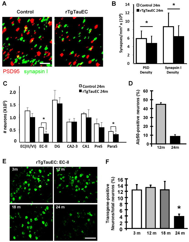
Neurofibrillary tangles advance from layer II of the entorhinal cortex (EC-II) toward limbic and association cortices as Alzheimer's disease evolves. However, the mechanism involved in this hierarchical pattern of disease progression is unknown. We describe a transgenic mouse model in which overexpression of human tau P301L is restricted to EC-II. Tau pathology progresses from EC transgene-expressing neurons to neurons without detectable transgene expression, first to EC neighboring cells, followed by propagation to neurons downstream in the synaptic circuit such as the dentate gyrus, CA fields of the hippocampus, and cingulate cortex. Human tau protein spreads to these regions and coaggregates with endogenous mouse tau. With age, synaptic degeneration occurs in the entorhinal target zone and EC neurons are lost. These data suggest that a sequence of progressive misfolding of tau proteins, circuit-based transfer to new cell populations, and deafferentation induced degeneration are part of a process of tau-induced neurodegeneration.


Figures

Figure 1
Figure 1. Medial entorhinal cortex-restricted expression of human P301L mutated tau causes progressive tauopathy in EC neurons
The rTgTauEC transgenic mice express human tauP301L under a tetracycline responsive promoter (tetO) and the tetracycline transactivator (tTA) under the neuropsin promoter (A). A low magnification view of a medial horizontal section of rTgTauEC mouse brain stained with DAPI (B) and fluorescent in situ hybridization (FISH) showing human tau mRNA (C) demonstrates the restriction of expression of human tauP301L to layer II of the medial entorhinal cortex (MEC) and the pre- and para-subiculum (Pre, Para). (A–C) The EC and MEC are outlined with a red dotted line. Note the lack of expression in the lateral entorhinal cortex (LEC), the subiculum (Sub), and the hippocampus including the dentate gyrus (DG), CA3 and CA1. There was no transgene expression in age matched controls (right panel, C). Scale bar, 1mm (B,C). (B) Higher magnification insets show the white outline EC area, Scale, 100μm. Immunohistolabeling with phosphorylation-independent tau antibody 5A6 shows that tau protein is intensely expressed from 3 months of age compared to an age-matched control brain (D, Scale bar, 1mm left, 200μm in higher magnification insets). Human tau expressing neurons in the MEC develop progressive tau pathology (E) beginning at 3 months of age with Alz50 positive tau misfolding in the axon terminal zone (molecular layer of the DG, left panel, scale bar 200 μm). By 6 months of age, MEC neurons have Alz50 positive tau staining in their soma, and the soma in this region increasingly accumulates pathological forms of tau with age, shown here by markers of later tau pathology including PHF1 (tau phosphorylated at Ser396 and Ser404) starting from 12 months, Gallyas silver stain positive neurons from 18 months, and Thioflavin S positive neurofibrillary tangles by 24 months of age. Scale bar 50 μm for all soma images.
Figure 2
Figure 2. Biochemical characterization of tau pathology reveals age dependent increase in tau hyperphosphorylation and presence of sarkosyl insoluble tau in rTgTauEC
(A) Representative immunoblots from western blots analysis of mouse brain extracts from 12, 18 and 24 month-old rTgTauEC and control mice. Antibodies specific to phospho tau [AT180 (pT231), PHF1 (pS396/404) and CP13 (pS202)], total tau, and human tau (htau) were used. Quantification of immunoblots shows that rTgTauEC mice present an age-dependent increase in total tau (B), human tau (results are expressed as percent of total tau expression at 24 months, the highest expression point defined as 100%) (C), and phospho epitopes (D–F) (n=4 mice per group). A series of ultracentrifugation and extraction steps were used to obtain sarkosyl-insoluble fractions of tau from whole brain of 24 month-old rTgTauEC, control (25 μg of protein loaded), and 18 month-old rTg4510 mice (5 μg of protein loaded) (positive control). Sarkosyl soluble (S) and insoluble fractions (I) were immunoblotted with total tau antibody (phosphorylation independent, Dako). Representative western blots of sarkosyl fractions are shown. (G). Control mice did not show sarkosyl insoluble tau. In contrast, immunoblotting of fractions from rTgTauEC mice revealed sarkosyl insoluble complexes of tau, of the same type as found in the brains of rTg4510 mice. A 64 kDa insoluble hyperphosphorylated tau species was detected by immunoblot in both rTg4510 and rTgTauEC brains, but were absent in age-matched control brains when analyzed using total tau antibody. In the soluble fraction, the 55 kDa species of tau were also present, similar to the species present in rTg4510 mice. (H) Shows quantification of samples of sarkosyl fractions (n=3 mice per group). Results are expressed as mean ± s.e.m. *P<0.05; **P<0.01; ***P<0.001.
Figure 3
Figure 3. Tau propagates through neural circuits to mRNA-negative cells
Several markers of pathological tau accumulation: Alz50 (A), PHF1 (B), and Gallyas silver staining (C), appear in brain regions synaptically connected to EC via the perforant pathway including DG, CA1, and CA2/3 (Scale bar 50 μm). FISH for tau mRNA (green), coupled with immunolabeling (red) with Alz50 (D) or HT7 (E) shows absence of mRNA in some cells containing the tau protein (arrows). Scale bar, 20 μm. Co-localization of tau mRNA and Alz50-positive aggregates was assessed by stereology at different ages, and compared to the total population of neurons carrying Alz50 aggregates. At 12 months of age, a third of the neurons affected by the tau pathology were expressing the transgene, while only ~3% were positive for MAPT gene at 24 months of age (F). FISH for MAPT mRNA (green), coupled with immunolabeling using HT7 (red) in the same EC sections from 17 month-old animals (E) was used to laser capture three different population of cells (G): 1) tau mRNA negative and human protein negative neurons; 2) mRNA negative and human tau protein positive neurons; 3) transgene mRNA positive and human tau protein positive neurons. (G) Total RNA was extracted and qPCR analysis of the cDNA product was carried out using primers against the transgenic human tau construct showing that the neurons which were human tau protein positive and were RNA negative by FISH did not express the tau transgene, confirming tau transmission to neurons that do not express the human tau transgene. Results are expressed as mean ± s.e.m. *P<0.001.
Figure 4
Figure 4. Human tau protein seeds endogenous mouse tau pathology
Immunohistochemistry using a mouse specific tau antibody (mTau) shows normal axonal distribution of mouse tau in the control mice and somatodendritic accumulation of mouse tau in rTgTauEC mice in the EC at 18 months of age (A). The mTau antibody showed no staining in tau knockout mice or human AD brain. Scale bars, 100 μm. Double labeling using Alz50 and mouse tau antibodies shows that Alz50 and mouse tau staining co-localize in neuronal cell bodies (B, Scale bar, 20 μm). (C) Whole brain extracts from rTgTauEC mice at different ages show an age-dependent increase in mouse tau. Representative blots are shown on top and quantification on the bottom (n=4 mice per group). (D) Sarkosyl insoluble and soluble fractions from whole brain of 18 and 24 month-old rTgTauEC probed using the mouse tau specific antibody mTau revealed that sarkosyl insoluble and soluble fractions both contain endogenous mouse tau (E). Immunoblotting of brain extracts using the mouse tau specific antibody confirmed its specificity since it does not recognize protein in tau knockout brain or human AD brain. Results are expressed as mean ± s.e.m. *P<0.05.
Figure 5
Figure 5. Axon terminals degenerate progressively in the terminal zone of EC-II axons
The terminal zone of axonal projections from the layer II MEC neurons with the granular layer (gl) and the middle- molecular layers (mml) which express the human tau transgene developed early accumulation of misfolded tau (Alz50 staining, A) which intensified up to 12 months of age. At 18 months, the terminal staining with Alz50 became fainter with DG neurons in the granular cell layer becoming more prominently stained. At 21 and 24 months, Alz50 staining in the terminal zone was patchy indicating degeneration of Alz50 containing axons (Scale bar 100 μm, A). Concomitant with this axonal degeneration, we observe increased microglial activation (Iba1 staining) at 24 months of age in rTgTauEC mice in the molecular layer of the dentate gyrus (B) Double Immunohistolabeling with Alz50 and Iba1 shows that the patches of Alz50 staining of the axon terminals in the middle molecular layer of dentate gyrus are surrounded by activated microglia (C, shown in higher magnification in the inset. Scale bars, 50 μm left panel), and 20 μm right panel). GFAP labeled astrocytes are more prevalent in rTgTauEC brain than controls (D, Scale bar, 50 μm). Co-localization of GFAP and PHF1-positive aggregates indicate uptake of tau by astrocytes (E, Scale bar, 20 μm).
Figure 6
Figure 6. Synaptic loss in the target zone of the perforant pathway and loss of EC-II neurons in rTgTauEC mice
Array tomography using synapsin I (green) and PSD95 (red) to label pre and postsynaptic structures in the middle molecular layer of the DG (A, scale bar 2 μm) shows pre and post-synaptic loss at 24 months of age in the middle molecular layer of the dentate gyrus (B), indicating loss of synapses between EC-II neurons and DG neurons. (C) Numbers of neurons were estimated by stereology for different areas of the brain (EC 3–6, layers III to VI of entorhinal cortex; EC 2, layer II; DG, granular layer of dentate gyrus; CA2-3; CA1; presubiculum; parasubiculum). Significant neuronal loss was detected at 24 months of age in the layer II of entorhinal cortex and parasubiculum, compared to age-matched control brain. (D) Similar stereological quantification showed that 47% of neurons in the EC were Alz50 positive at 12 months of age and approximately 10% at 24 months of age. (E and F) Levels of transgene expression: loss of neurons could not be explained by an increase in transgene expression with age since the level of transgene expression assessed by stereological counts of tau mRNA positive neurons labeled with in situ hybridization showed that the level of transgene expression in MEC did not change from 3 to 18 months of age and significantly decreased at 24 months (E, F Scale, 100μm). Results are expressed as mean ± s.e.m. *P<0.05.

Comment in

Similar articles

Cited by

References

    1. Ballatore C, Lee VM, Trojanowski JQ. Tau-mediated neurodegeneration in Alzheimer’s disease and related disorders. Nat Rev Neurosci. 2007;8:663–672. - PubMed
    1. Barten DM, Cadelina GW, Hoque N, DeCarr LB, Guss VL, Yang L, Sankaranarayanan S, Wes PD, Flynn ME, Meredith JE, Ahlijanian MK, Albright CF. Tau transgenic mice as models for cerebrospinal fluid tau biomarkers. J Alzheimers Dis. 2011;24:127–141. - PubMed
    1. Blennow K, Wallin A, Agren H, Spenger C, Siegfried J, Vanmechelen E. Tau protein in cerebrospinal fluid: a biochemical marker for axonal degeneration in Alzheimer disease? Mol Chem Neuropathol. 1995;26:231–245. - PubMed
    1. Braak H, Braak E. Neuropathological stageing of Alzheimer-related changes. Acta Neuropathol. 1991;82:239–259. - PubMed
    1. Canto CB, Wouterlood FG, Witter MP. What does the anatomical organization of the entorhinal cortex tell us? Neural Plast. 2008;2008:381243. - PMC - PubMed

Publication types

MeSH terms