. 2012:507:255-74.
doi: 10.1016/B978-0-12-386509-0.00013-2.

Gene delivery to the retina: from mouse to man

Affiliations

Gene delivery to the retina: from mouse to man

Jean Bennett et al. Methods Enzymol. 2012.

Abstract

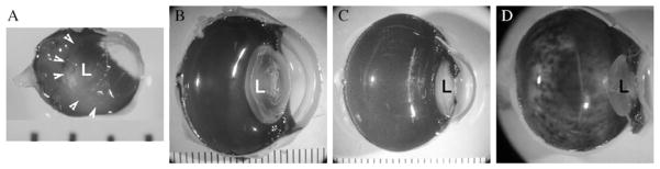
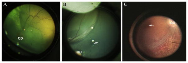
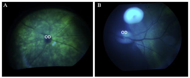
With the recent progress in identifying disease-causing genes in humans and in animal models, there are more and more opportunities for using retinal gene transfer to learn more about retinal physiology and also to develop therapies for blinding disorders. Success in preclinical studies for one form of inherited blindness have led to testing in human clinical trials. This paves the way to consider a number of other retinal diseases as ultimate gene therapy targets in human studies. The information presented here is designed to assist scientists and clinicians to use gene transfer to probe the biology of the retina and/or to move appropriate gene-based treatment studies from the bench to the clinic.


Figures

Figure 13.1
Figure 13.1
The space occupied by the lens relative to the vitreous cavity differs across species and dictates the surgical approach. In the mouse (A), the lens (L) occupies the majority of the cavity and so injections are usually carried out with a trans-choroidal approach. In larger animals ((B) dog, (C) monkey) and (D) humans, the lens is much smaller relative to the rest of the eye and so injections can be carried out from an anterior approach under direction visualization. Arrowheads in (A) indicate the borders of the lens. Distance between each bar in (A)–(C) is 1mm.
Figure 13.2
Figure 13.2
Appearance of the “bleb” immediately following subretinal injection in (A) dog, (B) non-human primate (NHP), and (C) human. OD, optic disc; arrow indicates the fovea in the NHP and the human. Panel (C) was taken from an intraoperative video recording.
Figure 13.3
Figure 13.3
Green fluorescent protein (GFP) is visible through illumination with blue light with an ophthalmoscope in animals after subretinal injection of a recombinant adeno-associated virus vector delivering the gene encoding GFP. The GFP is below the layer of the inner retinal vessels, which appear dark. (A) Mouse; (B) non-human primate; OD, optic disc.

References

    1. Acland GM, Aguirre GD, Ray J, et al. Gene therapy restores vision in a canine model of childhood blindness. Nat Genet. 2001;28:92–95. - PubMed
    1. Acland GM, Aguirre GD, Bennett J, et al. Long-term restoration of rod and cone vision by single dose rAAV-mediated gene transfer to the retina in a canine model of childhood blindness. Mol Ther. 2005;12:1072–1082. - PMC - PubMed
    1. Alexander JJ, Umino Y, Everhart D, et al. Restoration of cone vision in a mouse model of achromatopsia. Nat Med. 2007;13:685–687. - PMC - PubMed
    1. Ali R, Sarra GM, Stephens C, et al. Restoration of photoreceptor ultrastructure and function in retinal degeneration slow mice by gene therapy. Nat Genet. 2000;25:306–310. - PubMed
    1. Allocca M, Doria M, Petrillo M, et al. Serotype-dependent packaging of large genes in adeno-associated viral vectors results in effective gene delivery in mice. J Clin Invest. 2008;118:1955–1964. - PMC - PubMed

Publication types