

The targeted intracellular delivery of cytochrome C protein to tumors using lipid-apolipoprotein nanoparticles
- PMID: 22365810
- PMCID: PMC3307951
- DOI: 10.1016/j.biomaterials.2012.02.010
The targeted intracellular delivery of cytochrome C protein to tumors using lipid-apolipoprotein nanoparticles
Abstract
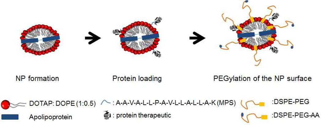
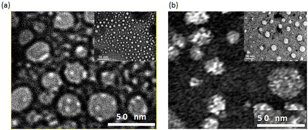
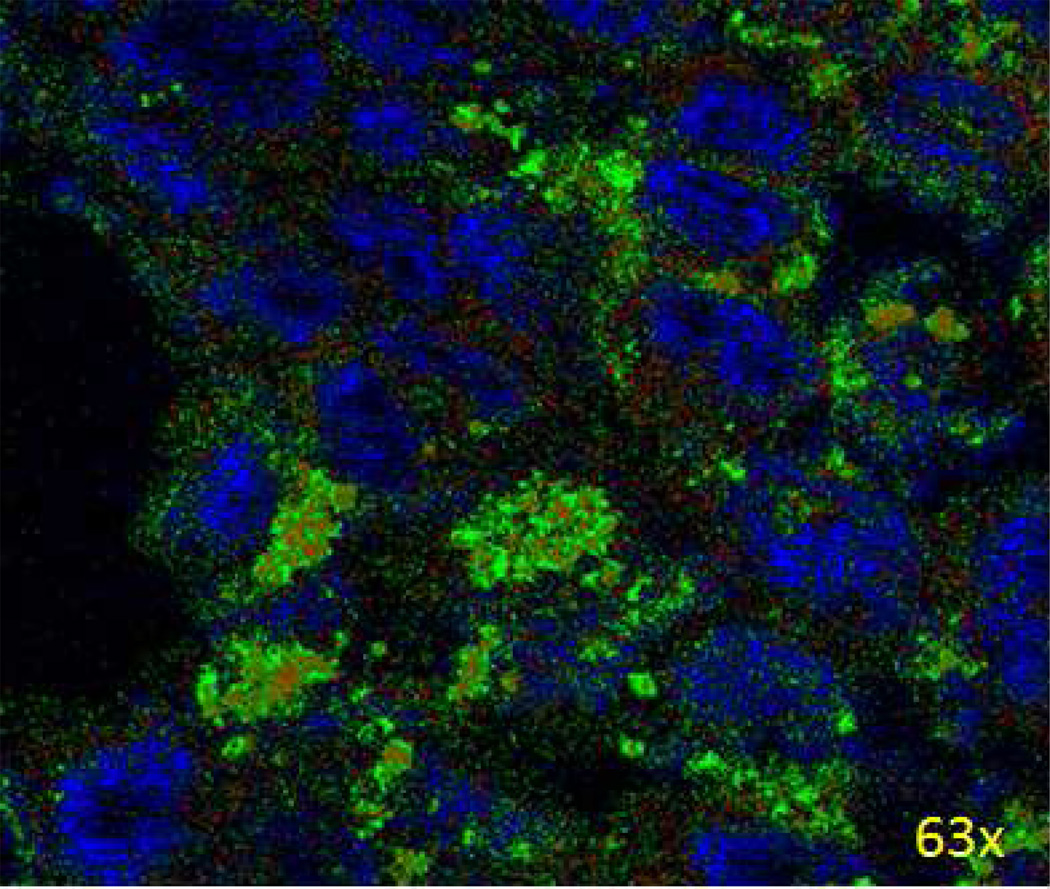
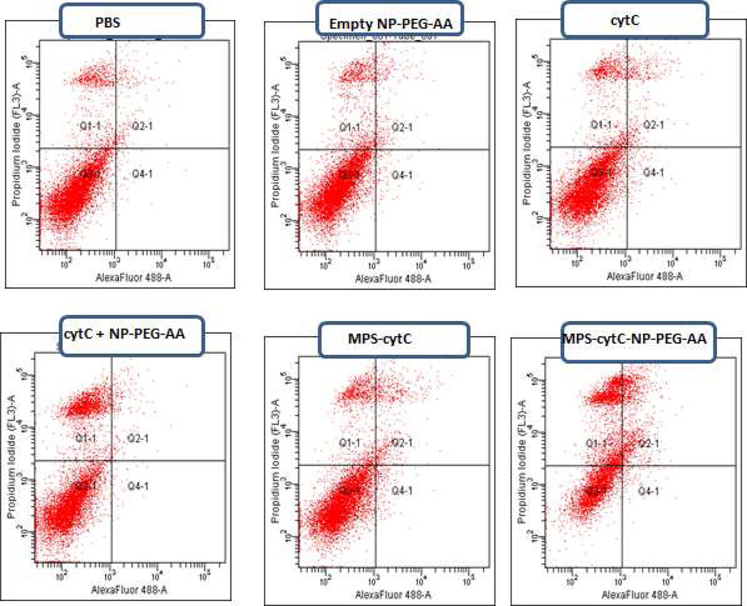
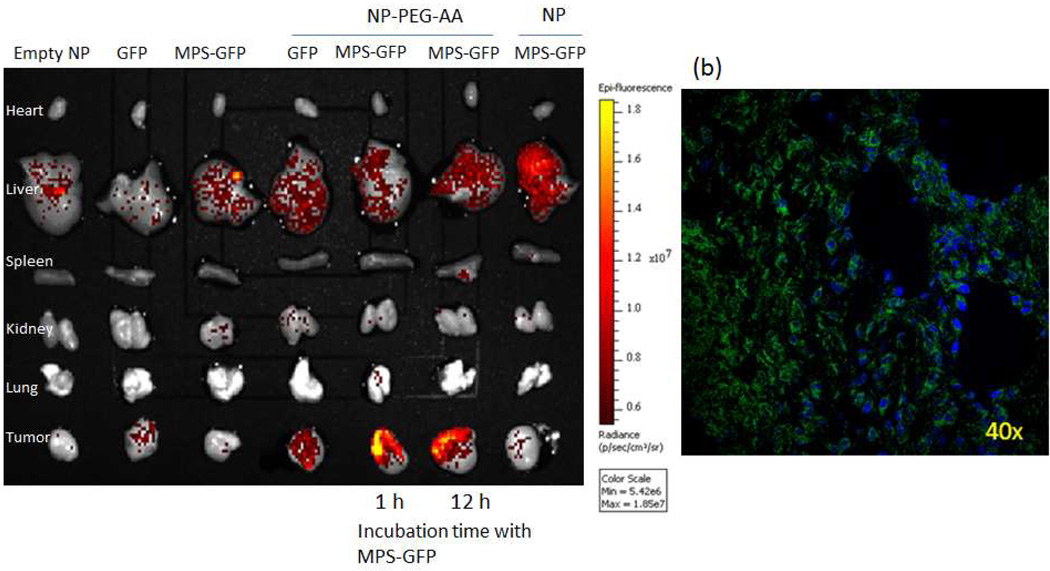
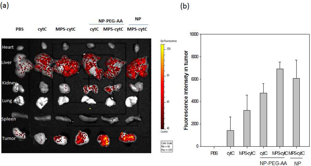
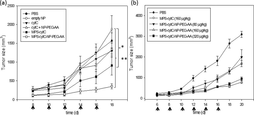
Intracellular-acting therapeutic proteins offer a promising clinical alternative to extracellular-acting agents, but are limited in efficacy by their low permeability into the cell cytoplasm. We have developed a nanoparticle (NP) composed of lipid (DOTAP/DOPE) and apolipoprotein (APOA-I) to mediate the targeted delivery of intracellular-acting protein drugs to non-small cell lung tumors. NPs were produced with either GFP, a fluorescent model protein, or cytochrome C (cytC), an inducer of apoptosis in cancer cells. GFP and cytC were separately conjugated with a membrane permeable sequence (MPS) peptide and were admixed with DOPE/DOTAP nanoparticle formulations to enable successful protein loading. Protein-loaded NPs were modified with DSPE-PEG-Anisamide to enable specific NP targeting to the tumor site in a xenograft model. The resulting particle was 20-30 nm in size and exhibited a 64-75% loading efficiency. H460 cells treated with the PEGylated MPS-cytC-NPs exhibited massive apoptosis. When MPS-GFP-NPs or MPS-cytC-NPs were intravenously administered in H460 tumor bearing mice, a specific tumor targeting effect with low NP accumulation in the liver was observed. In addition, MPS-cytC-NP treatment provoked a tumor growth retardation effect in H460 xenograft mice. We conclude that our NP enables targeted, efficacious therapeutic protein delivery for the treatment of lung cancer.
Copyright © 2012 Elsevier Ltd. All rights reserved.
Conflict of interest statement
The authors acknowledge that there was no conflict of interest or improper external influence on this work.
Figures

Similar articles
-
Turning an antiviral into an anticancer drug: nanoparticle delivery of acyclovir monophosphate.J Control Release. 2013 Sep 28;170(3):414-20. doi: 10.1016/j.jconrel.2013.06.009. Epub 2013 Jun 18. J Control Release. 2013. PMID: 23791977 Free PMC article.
-
Low density lipoprotein mimic nanoparticles composed of amphipathic hybrid peptides and lipids for tumor-targeted delivery of paclitaxel.Int J Nanomedicine. 2019 Sep 11;14:7431-7446. doi: 10.2147/IJN.S215080. eCollection 2019. Int J Nanomedicine. 2019. PMID: 31686815 Free PMC article.
-
Targeted delivery of EV peptide to tumor cell cytoplasm using lipid coated calcium carbonate nanoparticles.Cancer Lett. 2013 Jul 1;334(2):311-8. doi: 10.1016/j.canlet.2012.07.011. Epub 2012 Jul 14. Cancer Lett. 2013. PMID: 22796364 Free PMC article.
-
Biofunctionalized polymer-lipid supported mesoporous silica nanoparticles for release of chemotherapeutics in multidrug resistant cancer cells.Biomaterials. 2014 Apr;35(11):3650-65. doi: 10.1016/j.biomaterials.2014.01.013. Epub 2014 Jan 24. Biomaterials. 2014. PMID: 24462359
-
Lipid-polymer hybrid nanoparticles as a new generation therapeutic delivery platform: a review.Eur J Pharm Biopharm. 2013 Nov;85(3 Pt A):427-43. doi: 10.1016/j.ejpb.2013.07.002. Epub 2013 Jul 17. Eur J Pharm Biopharm. 2013. PMID: 23872180 Review.
Cited by
-
A small-molecule carrier for the intracellular delivery of a membrane-impermeable protein with retained bioactivity.Proc Natl Acad Sci U S A. 2024 Oct 29;121(44):e2407515121. doi: 10.1073/pnas.2407515121. Epub 2024 Oct 22. Proc Natl Acad Sci U S A. 2024. PMID: 39436658 Free PMC article.
-
Effect of size and pegylation of liposomes and peptide-based synthetic lipoproteins on tumor targeting.Nanomedicine. 2017 Aug;13(6):1869-1878. doi: 10.1016/j.nano.2017.04.009. Epub 2017 Apr 18. Nanomedicine. 2017. PMID: 28434931 Free PMC article.
-
Intracellular Protein Delivery: Approaches, Challenges, and Clinical Applications.BME Front. 2024 Jan 25;5:0035. doi: 10.34133/bmef.0035. eCollection 2024. BME Front. 2024. PMID: 38282957 Free PMC article. Review.
-
Engineering cytochrome-modified silica nanoparticles to induce programmed cell death.Chemistry. 2013 Dec 23;19(52):17891-8. doi: 10.1002/chem.201303239. Epub 2013 Nov 18. Chemistry. 2013. PMID: 24249039 Free PMC article.
-
Incorporation of histone derived recombinant protein for enhanced disassembly of core-membrane structured liposomal nanoparticles for efficient siRNA delivery.J Control Release. 2013 Nov 28;172(1):179-189. doi: 10.1016/j.jconrel.2013.08.015. Epub 2013 Aug 23. J Control Release. 2013. PMID: 23978682 Free PMC article.
References
-
- Tabata Y, Ikada Y. Protein release from gelatin matrices. Adv Drug Deliv Rev. 1998;31:287–301. - PubMed
-
- Manning M, Chou D, Murphy B, Payne R, Katayam D. Stability of protein pharmaceuticals: an update. Pharmaceutical Research. 2010;27:544–575. - PubMed
-
- Porjazoska A, Goracinova K, Mladenovska K, Glavas M, Simonovska M, Janjević EI, et al. Poly(lactide-co-glycolide) microparticles as systems for controlled release of proteins: preparation and characterization. Acta Pharm. 2004;54:215–229. - PubMed
-
- Panyam J, Dali MM, Sahoo SK, Ma W, Chakravarthi SS, Amidon GL, et al. Polymer degradation and in vitro release of a model protein from poly(D, L lactide-co-glycolide) nano- and microparticles. J Control Release. 2003;92:173–187. - PubMed
-
- Mohamed F, Van der Walle CF. Engineering biodegradable polyester particles with specific drug targeting and drug release properties. J Pharm Sci. 2008;97:71–87. - PubMed
Publication types
MeSH terms
Substances
Grants and funding
LinkOut - more resources
Full Text Sources
Miscellaneous
