. 2012 Mar 30;149(1):188-201.
doi: 10.1016/j.cell.2012.01.046. Epub 2012 Feb 23.

Young dentate granule cells mediate pattern separation, whereas old granule cells facilitate pattern completion

Affiliations

Young dentate granule cells mediate pattern separation, whereas old granule cells facilitate pattern completion

Toshiaki Nakashiba et al. Cell. .

Abstract

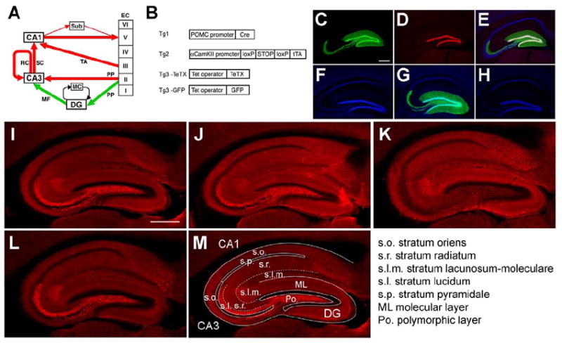
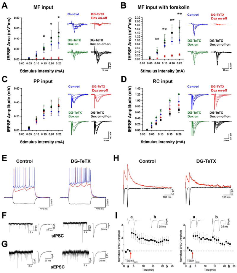
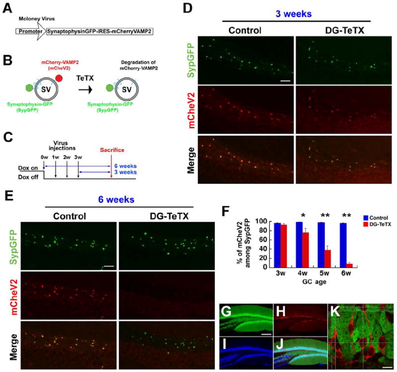
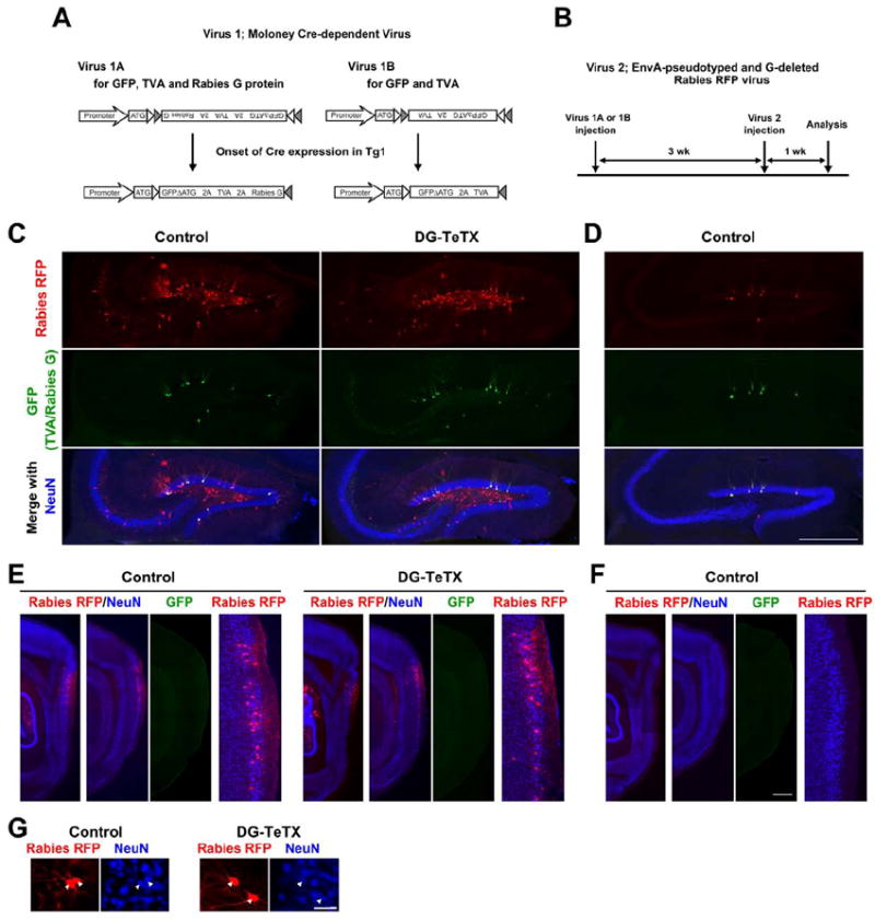
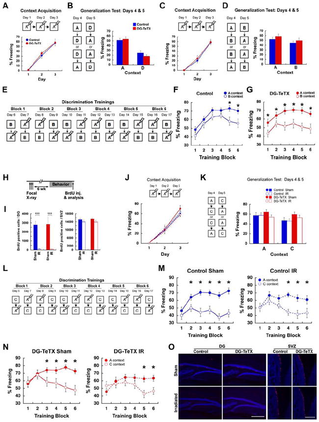
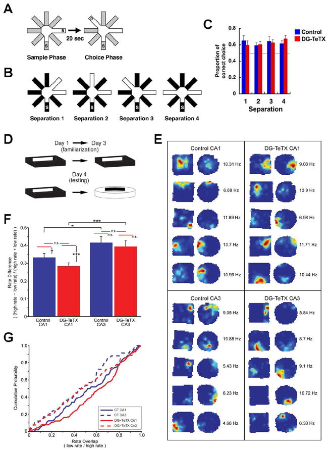
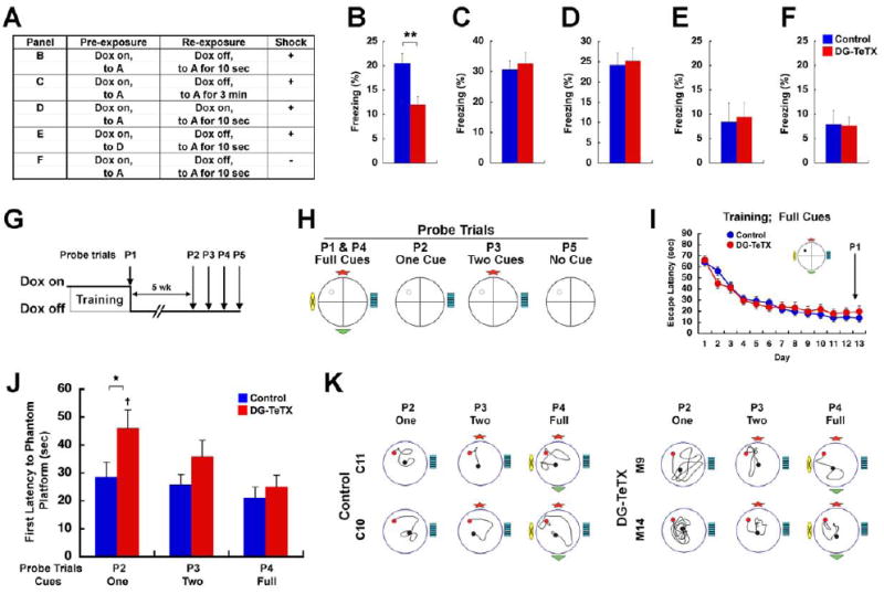
Adult-born granule cells (GCs), a minor population of cells in the hippocampal dentate gyrus, are highly active during the first few weeks after functional integration into the neuronal network, distinguishing them from less active, older adult-born GCs and the major population of dentate GCs generated developmentally. To ascertain whether young and old GCs perform distinct memory functions, we created a transgenic mouse in which output of old GCs was specifically inhibited while leaving a substantial portion of young GCs intact. These mice exhibited enhanced or normal pattern separation between similar contexts, which was reduced following ablation of young GCs. Furthermore, these mutant mice exhibited deficits in rapid pattern completion. Therefore, pattern separation requires adult-born young GCs but not old GCs, and older GCs contribute to the rapid recall by pattern completion. Our data suggest that as adult-born GCs age, their function switches from pattern separation to rapid pattern completion.


Figures

Figure 1
Figure 1. Application of the DICE-K method to the MF pathway
(A) Excitatory pathways in the hippocampus and EC. Red and green colors designate alternative pathways. (B) Transgenic constructs. For the production of the DG-TeTX mouse and the model mouse (DG-GFP), Tg1, Tg2 and Tg3-TeTX mice and Tg1, Tg2 and Tg3-GFP mice were crossed, respectively. (C to E) A hippocampal section from a DG-GFP mouse raised on Dox, followed by a 2-week Dox withdrawal, co-stained with anti-GFP (green), anti-Prox1 [a marker for DG GCs (red)] and anti-NeuN [a marker for neurons (blue)]. Images through a green filter (C), through a red filter (D) and through all three filters (E). (F to H) Hippocampal sections co-stained with anti-GFP (green) and DAPI [a marker for cell nuclei (blue)] from a chronically repressed (Dox-on) DG-GFP mouse (F), from a mouse after a 2-week Dox withdrawal (G) and from a mouse in which withdrawal was followed by a 3-week Dox readministration (H). (I to L) Hippocampal sections stained with anti-VAMP2 from control (I) and DG-TeTX mice (J) both raised on Dox; from a DG-TeTX mouse raised on Dox, followed by a 4-week Dox withdrawal (K) and further followed by a 4-week Dox readministration (L). (M) Locations of various hippocampal strata. Scale bars, 500 μm. See also Figure S1.
Figure 2
Figure 2. Blockade of MF synaptic transmission in DG-TeTX mice and comparison of firing and synaptic properties of young adult-born GCs in control and activated DG-TeTX mice
(A and B) Input-output (I-O) relationships of MF input to CA3 in control ACSF (A) and in the presence of forskolin (B). (C) I-O relationships of the PP input to CA3. (D) I-O relationships of the RC input to CA3. Blue, control littermates (Tg1xTg3-TeTX) raised under Dox-on conditions, followed by a 6-week Dox withdrawal; green, repressed DG-TeTX mice (always Dox-on); red, activated DG-TeTX mice raised under Dox-on conditions, followed by a 6-week Dox withdrawal; black, DG-TeTX mice raised under Dox-on conditions, followed by a 6-week Dox withdrawal, followed by a 4-week Dox readministration. For all I-O relations shown, n=4-7 from at least four different mice per genotype/condition. Representative traces for each condition and pathway are indicated on the right. *p<0.05, **p<0.01 between control and activated DG-TeTX mice (t-test). (E to I) Firing and synaptic properties of young (3- to 4-week-old) adult-born GCs in control and activated DG-TeTX mice. (E) Representative traces showing membrane and firing properties of young GCs to hyperpolarizing (-50 pA; black traces) and depolarizing (+50 and +100 pA, red and blue traces, respectively) current injections. (F and G)Representative gap-free traces of pharmacologically isolated spontaneous GABAA and AMPA receptor-mediated IPSCs (F) and EPSCs (G) with ensemble averages (right traces) in young GCs. (H) Paired pulse (50 ms inter-stimulus interval) PP-evoked EPSCs at a holding potential of -70mV (black traces, AMPAR-mediated current) and at a holding potential of +40 mV (red traces, NMDAR-mediated current) in representative recordings from young GCs. (I) Pooled data showing long-term potentiation of PP-evoked AMPAR-mediated EPSCs in response to a theta burst induction protocol (see Experimental Procedures) in young GCs in control (n=8) and activated DG-TeTX mice (n=6). Inset traces a and b show EPSCs taken at the time indicated on the x-axes. Data represent mean ± SEM. See also Figure S2 and Table S1.
Figure 3
Figure 3. Integrity of MF transmission from young adult-born GCs
(A) A Moloney viral vector encoding bicistronic SypGFP and mCheV2. (B) SypGFP- and mCheV2-labeled synaptic vesicles (SV). TeTX cleaves the mCheV2, leading to the loss of mCheV2 immunoreactivity. (C) Dox diet, viral injection and mouse sacrifice schedules. (D and E) Presence of VAMP2 at MF boutons from 3-week-old adult-born GCs (D) and its absence at MF boutons from 6-week-old adult-born GCs (E). S. lucidum areas of hippocampal sections stained with anti-GFP (green) and anti-mCherry (red) from control and activated DG-TeTX mice. (F) Proportion of mCheV2-positive puncta among sypGFP-positive puncta at various GC ages. At least three different mice per genotype/condition were used. *p<0.05 for 4-week-old GCs; **p<0.01 for 5- and 6-week-old GCs (t-test). (G to K) A hippocampal section from a DG-GFP mouse co-stained with anti-GFP (green, G), anti-DCX (red, H) and anti-NeuN (blue, I; merge, J). (K) A confocal image at a single z-axis with green and red filters. Optical sections along the horizontal or vertical lines across multiple z-axes are shown on the top and left, respectively. Scale bar in D and E, 25 μm; G-J, 250 μm; K, 10 μm. Data represent mean ± SEM. See also Figure S3.
Figure 4
Figure 4. Young adult-born GCs receive synaptic inputs
(A) Two different Cre-dependent Moloney viral vectors. Virus 1A (left) expresses GFP, TVA (a receptor for EnvA) and rabies G glycoprotein, whereas Virus 1B (right) expresses GFP and TVA. Cre-loxP recombination by Tg1 occurs between 1 and 2 weeks after the Moloney virus injection (Figure S4). (B) Schedule of analysis relative to viral injections. (C) Hippocampal sections from control and activated DG-TeTX mice injected with Virus 1A and Virus 2 co-stained with anti-RFP (red), anti-GFP (green) and anti-NeuN (blue). (D) A hippocampal section from a control mouse injected with Virus 1B and Virus 2. (E) Parasagittal sections at two different medio-lateral levels covering the EC (first and second columns) from control and activated DG-TeTX mice injected with Virus 1A and Virus 2. GFP images (third column) taken from sections shown in the second column. High magnification images of the EC superficial layers (fourth column) are from images in the second column. (F) Parasagittal sections from a control mouse injected with Virus 1B and Virus 2. (G) RFP- and NeuN-positive cells (arrowheads) in the EC. Scale bars in C-F, 500 μm; G, 50 μm. See also Figure S4.
Figure 5
Figure 5. DG-TeTX mice show enhanced contextual discrimination in a highly similar context pair and their intact young GCs are necessary for this discrimination
(A and B) Contextual discrimination between a very distinct context pair, A and D. (A) Freezing levels during the acquisition (blue, control; red, DG-TeTX; n=12 per genotype). (B) Freezing levels in A and D during the generalization test. (C to G) Another set of mice (n=12 per genotype) were subjected to contextual discrimination between a very similar context pair, A and B. (C) Freezing levels during the acquisition. (D) Freezing levels in A and B during the generalization test. (E) Experimental procedure for discrimination training between A and B. (F and G) Freezing levels in A (filled circles) and B (open circles) of control (blue, F) and DG-TeTX mice (red, G). (H to O) Activated DG-TeTX and control littermates with either IR or Sham (n=14 per group) were subjected to contextual discrimination between a slightly distinct context pair, A and C. (H) Mice were focally irradiated 6 weeks prior to contextual discrimination fear conditioning. (I) The number of BrdU-positive cells in the DG and in the subventricular zone (SVZ; blue, control; red, DG-TeTX; ***p<0.001 in both genotypes, t-test). (J) Freezing levels during the acquisition (blue, control; red, DG-TeTX; filled circles and solid lines, Sham; open circles and broken lines, IR). (K) Freezing levels in A and C during the generalization test. (L) Experimental procedure for discrimination trainings between A and C. (M and N) Freezing levels in A (filled circles) and C (open circles) of control (M) and DG-TeTX mice (N). (O) DG and SVZ area of sections stained with anti-BrdU (red) and anti-NeuN (blue). * indicates Scheffé’s correction for multiple comparison, p<0.05. Scale bars in O, 500 μm. Data represent mean ± SEM. See also Figure S5.
Figure 6
Figure 6. Normal spatial discrimination and normal rate remapping in DG-TeTX mice
(A) Pattern separation was tested using a DNMP protocol by varying angles between sample and reward arms (S, start arm; R, reward; see Experimental Procedures). (B) Two arms were separated by 45°, 90°, 135° and 180° in separation 1-4, respectively. (C) Proportions of correct choices as a function of arm separation angles. The horizontal gray line represents chance. n=12 per genotype. (D) Example of behavioral pattern separation paradigm. (E) Place field examples from control (left) and DG-TeTX mice (right) for CA1 (top) and CA3 (bottom) pyramidal cells. The two maps from each example were normalized to the maximum peak firing rate between the maps, indicated to the right of each pair. (F) Rate remapping index for place cells recorded from CA1 (left) and CA3 (right) regions of control (blue) and DG-TeTX mice (red). There was a similar significant difference in rate between CA1 and CA3 in control and DG-TeTX mice (*p<0.05 and ***p<0.001, respectively, t-test). The red bars above each column represent the estimated rate difference (ERD) expected given independent firing in the two boxes for that region/genotype. Only significant differences were observed between the actual and ERD in the CA1 regions of control and DG-TeTX mice (*p=0.012 and ***p<0.001, respectively, t-test). (G) Cumulative probability histograms of the overlap values (low rate/high rate) for each genotype and region. Data represent mean ± SEM. See also Table S2.
Figure 7
Figure 7. DG-TeTX mice exhibit deficits in pattern completion-mediated contextual and spatial recall
(A to F) Pre-exposure-dependent contextual fear conditioning. (A) Experimental procedure for B to F. Repressed DG-TeTX and control littermates were subjected to a pre-exposure session (for 5 consecutive days, 10 min per day) in either context A or D. The mice were then shifted to a Dox-free diet (except in D). Next, the mice were (re)-exposed to context A and then received a single footshock within either 10 s or 3 min. Mice in F did not receive a footshock. (B to F) Freezing levels in context A measured 1 day after (re)-exposure (blue, control; red, DG-TeTX). (n=35 and 36 in B for DG-TeTX mice and control mice, respectively; n=24, 24, 12 and 12 for both genotypes in C, D, E and F, respectively; **p<0.01, t-test). (G to K) Spatial memory recall with various cue conditions in the standard Morris water maze task. (G) Experimental procedure. Repressed DG-TeTX (n=23) and control littermates (n=24) were trained to locate a hidden platform location in the full-cue condition. The mice were then switched to a Dox-free diet. The P1 probe trial was conducted under the Dox-on condition, whereas the P2-P5 probe trials were conducted under the Dox-off condition. (H) Probe trials with cue manipulation. (I) Escape latencies to the hidden platform during the training (blue, control; red, DG-TeTX). (J) First latencies to the phantom platform during the probe trials. (K) Examples of the swim path to the phantom platform (red) for the probe trials (the black circle represents the start point). *p<0.05; p<0.01 in DG-TeTX mice, P2 vs. P4. Data represent mean ± SEM. See also Figure S6 and S7.

Comment in

Similar articles

Cited by

References

    1. Altman J, Bayer SA. Migration and distribution of two populations of hippocampal granule cell precursors during the perinatal and postnatal periods. J Comp Neurol. 1990;301:365–381. - PubMed
    1. Altman J, Das GD. Autoradiographic and histological evidence of postnatal hippocampal neurogenesis in rats. J Comp Neurol. 1965;124:319–335. - PubMed
    1. Amaral DG, Ishizuka N, Claiborne B. Neurons, numbers and the hippocampal network. Prog Brain Res. 1990;83:1–11. - PubMed
    1. Bakker A, Kirwan CB, Miller M, Stark CEL. Pattern separation in the human hippocampal CA3 and dentate gyrus. Science. 2008;319:1640–1642. - PMC - PubMed
    1. Braz JM, Rico B, Basbaum AI. Transneuronal tracing of diverse CNS circuits by Cre-mediated induction of wheat germ agglutinin in transgenic mice. PNAS U S A. 2002;99:15148–15153. - PMC - PubMed

Publication types

Substances