. 2012 Nov;70(11):2656-68.
doi: 10.1016/j.joms.2011.12.030. Epub 2012 Feb 25.

Inductive, scaffold-based, regenerative medicine approach to reconstruction of the temporomandibular joint disk

Affiliations

Inductive, scaffold-based, regenerative medicine approach to reconstruction of the temporomandibular joint disk

Bryan N Brown et al. J Oral Maxillofac Surg. 2012 Nov.

Abstract

Purpose: A device composed of extracellular matrix (ECM) was investigated as an inductive template in vivo for reconstruction of the temporomandibular joint (TMJ) disk after discectomy.

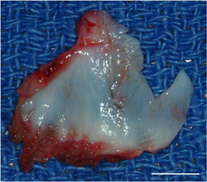
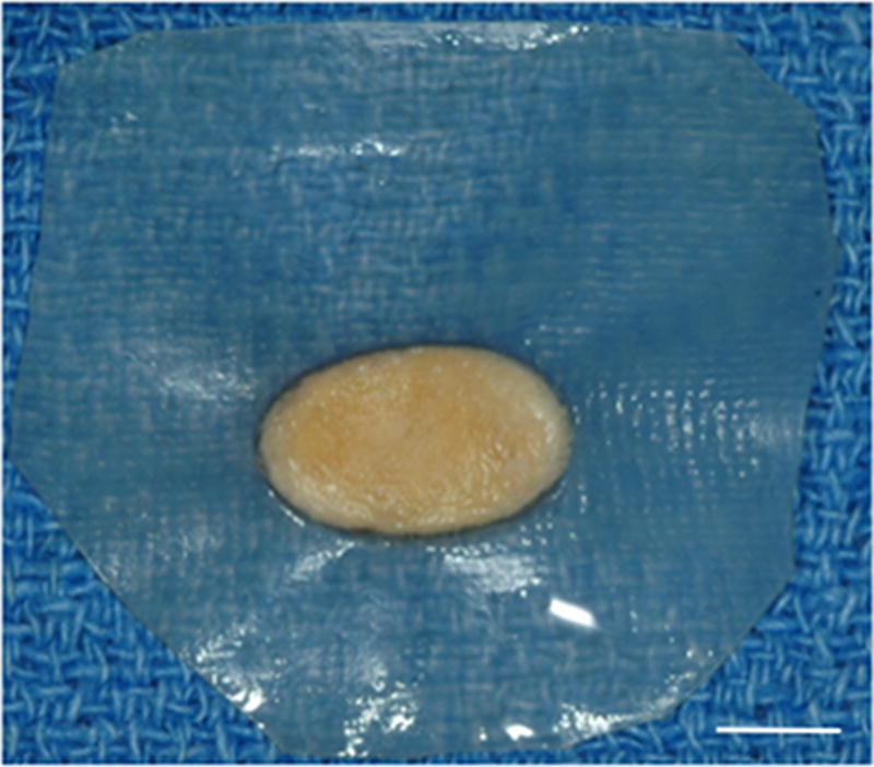
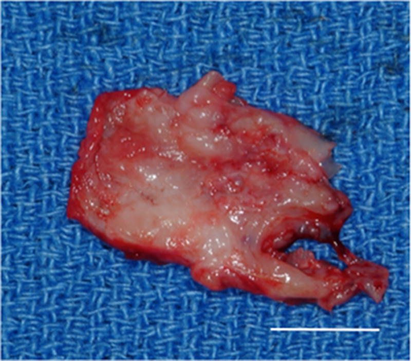
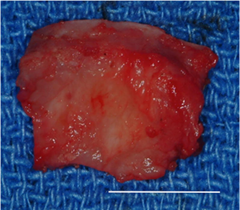
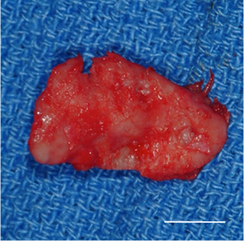
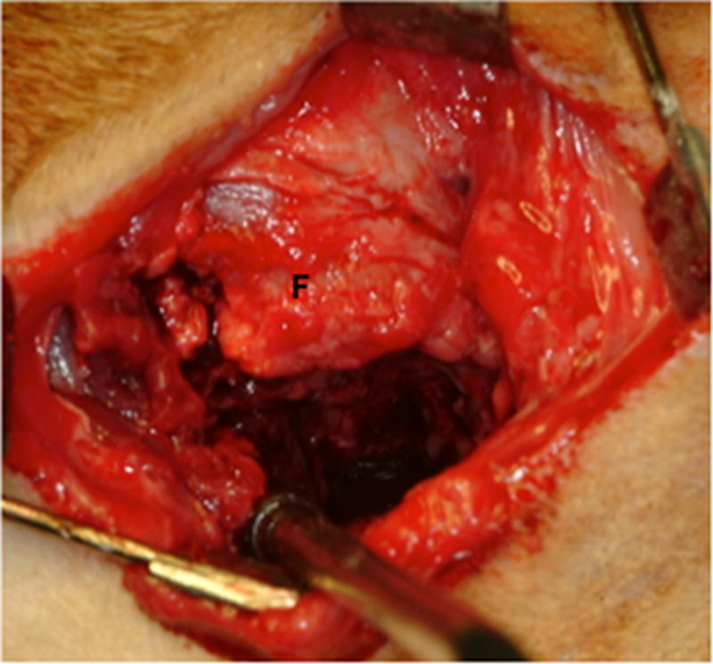
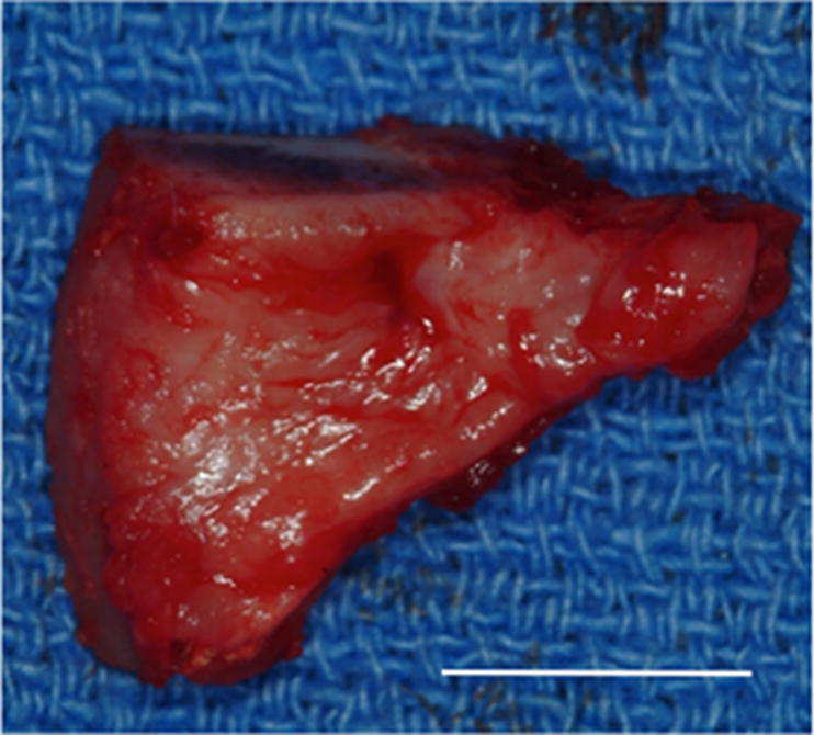
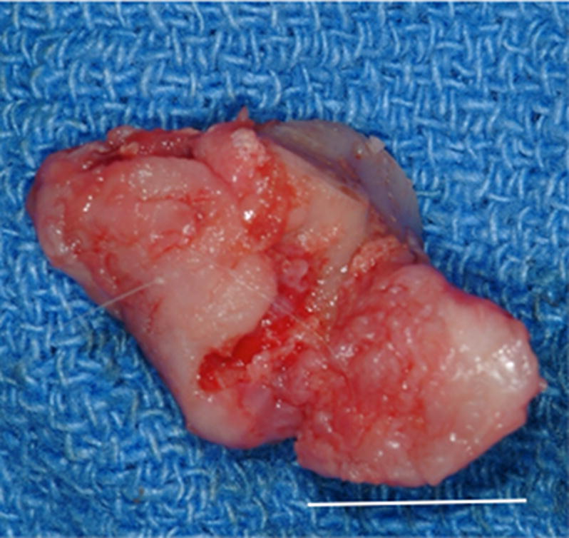
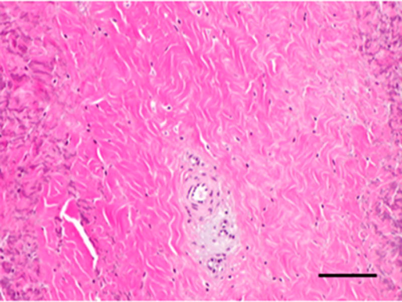
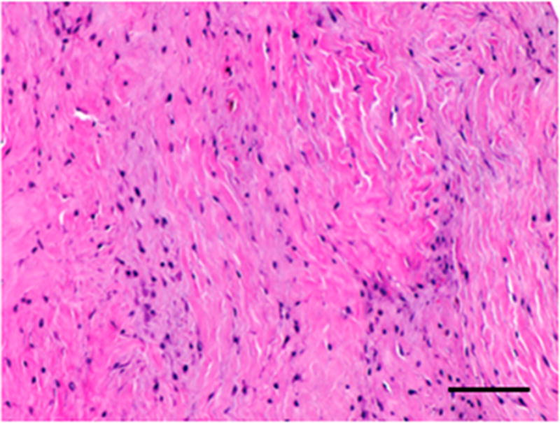
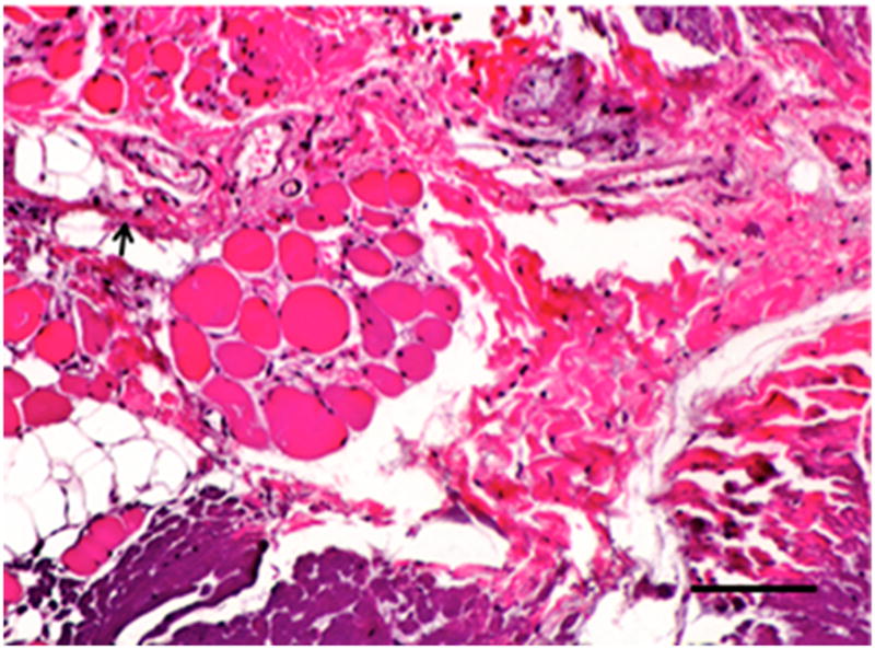
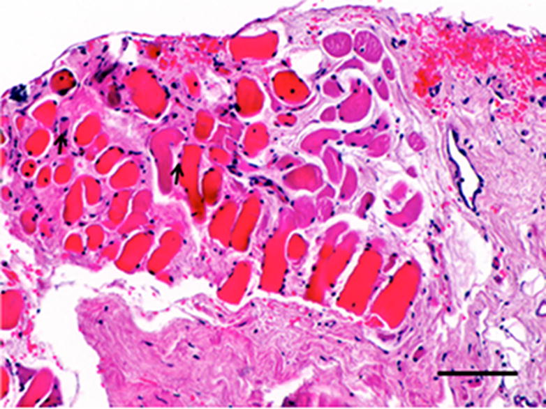
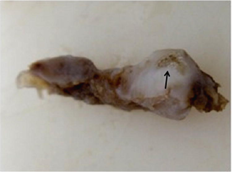
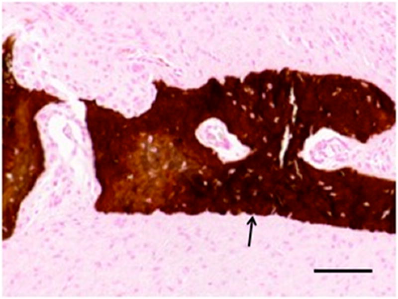
Materials and methods: A scaffold material composed of porcine-derived ECM was configured to mimic the shape and size of the TMJ. This device was implanted in a canine model of bilateral TMJ discectomy. After discectomy, 1 side was repaired with an ECM scaffold material and the contralateral side was left empty as a control. At 6 months after implantation, the joint space was opened, the joints were evaluated for signs of gross pathologic degenerative changes, and newly formed tissue was excised for histologic, biochemical, and biomechanical analysis.

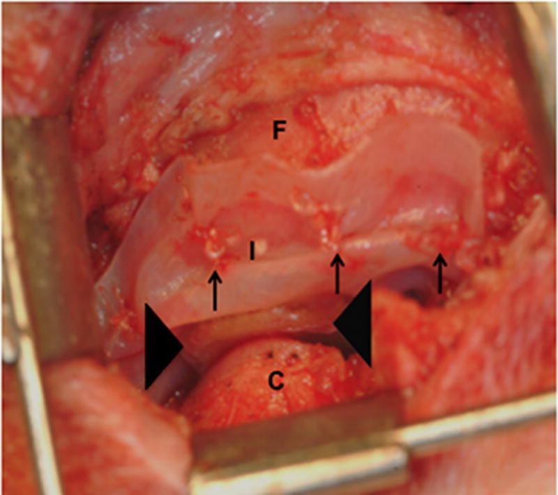
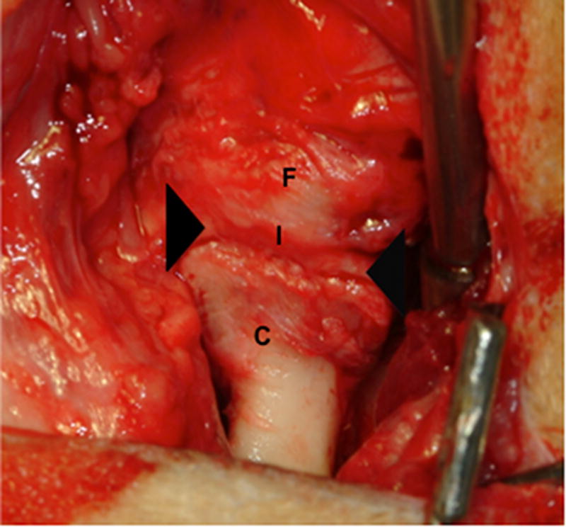
Results: The results showed that implantation of an initially acellular material supported the formation of site-appropriate, functional host tissue that resembled that of the native TMJ disk. Furthermore, this prevented gross degenerative changes in the temporal fossa and mandibular condyle. No tissue formation and mild to severe gross pathologic changes were observed in the contralateral controls.

Conclusions: These results suggest that an ECM-based bioscaffold could represent an off-the-shelf solution for TMJ disk replacement.


Figures

Figure 1
Figure 1
Images showing the surgical procedure. The native disk (A) is exposed and excised leaving the joint space empty. UBM implants (B) were hydrated and trimmed to size prior to placement between the condyle and fossa on the experimental side and attachment through the fossa (C). Note sutures through the anchoring site (arrows) and interpositional pillow-like core (arrowheads). The contralateral side was left devoid of a meniscal substitute. C = condyle, F = fossa, M = disk, and I = implanted UBM-ECM device. Scale bars in A and B = 5 mm.
Figure 1
Figure 1
Images showing the surgical procedure. The native disk (A) is exposed and excised leaving the joint space empty. UBM implants (B) were hydrated and trimmed to size prior to placement between the condyle and fossa on the experimental side and attachment through the fossa (C). Note sutures through the anchoring site (arrows) and interpositional pillow-like core (arrowheads). The contralateral side was left devoid of a meniscal substitute. C = condyle, F = fossa, M = disk, and I = implanted UBM-ECM device. Scale bars in A and B = 5 mm.
Figure 2
Figure 2
Images showing the gross morphologic appearance of the TMJ space at six months post-surgery. The experimental side (A) was characterized by the presence of interpositional material between the articulating surfaces of the fossa and condyle. Upon removal of the condyle, the newly formed tissue was removed and retained for histologic and mechanical testing. The newly formed tissue (B) is shown and was characterized by a white shiny appearance resembling that of the native tissue. The articulating portions of the fossa (C) and condyle (D) were characterized by a grossly normal looking articulating surface. No interpositional tissue formation was observed on the control sides. Upon removal of the condyle, mild to severe (E) degenerative changes in the surface of the fossa could be observed and were accompanied by the formation of varying degrees of granulation tissue and adhesions within the joint space. The excised fossa (F) and condylar (G) tissues were characterized by mild to severe gross pathologic changes in the articulating surface. C = condyle, F = fossa, I = newly formed interpositional tissue. Black arrowheads denote the location of the observed interpositional material in image A. Scale bars = 10 mm.
Figure 2
Figure 2
Images showing the gross morphologic appearance of the TMJ space at six months post-surgery. The experimental side (A) was characterized by the presence of interpositional material between the articulating surfaces of the fossa and condyle. Upon removal of the condyle, the newly formed tissue was removed and retained for histologic and mechanical testing. The newly formed tissue (B) is shown and was characterized by a white shiny appearance resembling that of the native tissue. The articulating portions of the fossa (C) and condyle (D) were characterized by a grossly normal looking articulating surface. No interpositional tissue formation was observed on the control sides. Upon removal of the condyle, mild to severe (E) degenerative changes in the surface of the fossa could be observed and were accompanied by the formation of varying degrees of granulation tissue and adhesions within the joint space. The excised fossa (F) and condylar (G) tissues were characterized by mild to severe gross pathologic changes in the articulating surface. C = condyle, F = fossa, I = newly formed interpositional tissue. Black arrowheads denote the location of the observed interpositional material in image A. Scale bars = 10 mm.
Figure 3
Figure 3
Histopathologic and immunolabeling findings. Microscopic views of hematoxylin and eosin staining of the bulk of the native (A) and remodeled tissues (B) showing a highly aligned collagenous matrix and spindle shaped cells. Microscopic views of hematoxylin and eosin staining of the peripheral muscular attachments of the native (C) and remodeled (D) tissues showing bundles of skeletal muscle within highly aligned collagenous matrix. CD31 and SMA labeling was observed only around the vascular structures observed primarily at the periphery of the tissues (not shown). All images are 20X original magnification.. Scale bar = 100 μm.
Figure 3
Figure 3
Histopathologic and immunolabeling findings. Microscopic views of hematoxylin and eosin staining of the bulk of the native (A) and remodeled tissues (B) showing a highly aligned collagenous matrix and spindle shaped cells. Microscopic views of hematoxylin and eosin staining of the peripheral muscular attachments of the native (C) and remodeled (D) tissues showing bundles of skeletal muscle within highly aligned collagenous matrix. CD31 and SMA labeling was observed only around the vascular structures observed primarily at the periphery of the tissues (not shown). All images are 20X original magnification.. Scale bar = 100 μm.
Figure 4
Figure 4
Area of calcification observed in remodeled tissues excised from one animal. Grossly identifiable calcification was observed outside of the area of articulation within the remodeled tissues in one animal (A). von Kossa staining was used to confirm the formation of calcified tissue (B). Calcification was not observed in other animals. Arrow denotes area of calcified tissue formation in A. Image B is 20X original magnification. Scale bar = 100 μm.
Figure 4
Figure 4
Area of calcification observed in remodeled tissues excised from one animal. Grossly identifiable calcification was observed outside of the area of articulation within the remodeled tissues in one animal (A). von Kossa staining was used to confirm the formation of calcified tissue (B). Calcification was not observed in other animals. Arrow denotes area of calcified tissue formation in A. Image B is 20X original magnification. Scale bar = 100 μm.

References

    1. Alonso A, Kaimal S, Look J, Swift J, Fricton J, Myers S, et al. A quantitative evaluation of inflammatory cells in human temporomandibular joint tissues from patients with and without implants. J Oral Maxillofac Surg. 2009;67:788–96. - PubMed
    1. Ferreira JN, Ko CC, Myers S, Swift J, Fricton JR. Evaluation of surgically retrieved temporomandibular joint alloplastic implants: pilot study. J Oral Maxillofac Surg. 2008;66:1112–24. - PMC - PubMed
    1. Fricton JR, Look JO, Schiffman E, Swift J. Long-term study of temporomandibular joint surgery with alloplastic implants compared with nonimplant surgery and nonsurgcal rehabilitation for painful temporomandibular joint disc displacement. J Oral Maxillofac Surg. 2002;60:1400–11. discussion 11-2. - PubMed
    1. Dolwick MF, Aufdemorte TB. Silicone-induced foreign body reaction and lymphadenopathy after temporomandibular join arthroplasty. Oal Surg Oral Medtroral Pathol. 1985;59:449–52. - PubMed
    1. Dimitroulis G. A critical review of interpositional grafts following temporomandibular joint discectomy with an overview of the dermis-fat graft. Int J Oral Maxillofac Surg. 2011;40:561–8. - PubMed

Publication types