Presence of a large β(1-3)glucan linked to chitin at the Saccharomyces cerevisiae mother-bud neck suggests involvement in localized growth control
- PMID: 22366124
- PMCID: PMC3318300
- DOI: 10.1128/EC.05328-11
Presence of a large β(1-3)glucan linked to chitin at the Saccharomyces cerevisiae mother-bud neck suggests involvement in localized growth control
Abstract
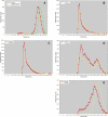
Previous results suggested that the chitin ring present at the yeast mother-bud neck, which is linked specifically to the nonreducing ends of β(1-3)glucan, may help to suppress cell wall growth at the neck by competing with β(1-6)glucan and thereby with mannoproteins for their attachment to the same sites. Here we explored whether the linkage of chitin to β(1-3)glucan may also prevent the remodeling of this polysaccharide that would be necessary for cell wall growth. By a novel mild procedure, β(1-3)glucan was isolated from cell walls, solubilized by carboxymethylation, and fractionated by size exclusion chromatography, giving rise to a very high-molecular-weight peak and to highly polydisperse material. The latter material, soluble in alkali, may correspond to glucan being remodeled, whereas the large-size fraction would be the final cross-linked structural product. In fact, the β(1-3)glucan of buds, where growth occurs, is solubilized by alkali. A gas1 mutant with an expected defect in glucan elongation showed a large increase in the polydisperse fraction. By a procedure involving sodium hydroxide treatment, carboxymethylation, fractionation by affinity chromatography on wheat germ agglutinin-agarose, and fractionation by size chromatography on Sephacryl columns, it was shown that the β(1-3)glucan attached to chitin consists mostly of high-molecular-weight material. Therefore, it appears that linkage to chitin results in a polysaccharide that cannot be further remodeled and does not contribute to growth at the neck. In the course of these experiments, the new finding was made that part of the chitin forms a noncovalent complex with β(1-3)glucan.
Figures


Similar articles
-
Two novel techniques for determination of polysaccharide cross-links show that Crh1p and Crh2p attach chitin to both beta(1-6)- and beta(1-3)glucan in the Saccharomyces cerevisiae cell wall.Eukaryot Cell. 2009 Nov;8(11):1626-36. doi: 10.1128/EC.00228-09. Epub 2009 Sep 4. Eukaryot Cell. 2009. PMID: 19734368 Free PMC article.
-
Altered extent of cross-linking of beta1,6-glucosylated mannoproteins to chitin in Saccharomyces cerevisiae mutants with reduced cell wall beta1,3-glucan content.J Bacteriol. 1997 Oct;179(20):6279-84. doi: 10.1128/jb.179.20.6279-6284.1997. J Bacteriol. 1997. PMID: 9335273 Free PMC article.
-
Architecture of the yeast cell wall. Beta(1-->6)-glucan interconnects mannoprotein, beta(1-->)3-glucan, and chitin.J Biol Chem. 1997 Jul 11;272(28):17762-75. doi: 10.1074/jbc.272.28.17762. J Biol Chem. 1997. PMID: 9211929
-
Biosynthesis of cell wall and septum during yeast growth.Arch Med Res. 1993 Autumn;24(3):301-3. Arch Med Res. 1993. PMID: 8298281 Review.
-
beta-1,6-Glucan synthesis in Saccharomyces cerevisiae.Mol Microbiol. 2000 Feb;35(3):477-89. doi: 10.1046/j.1365-2958.2000.01713.x. Mol Microbiol. 2000. PMID: 10672173 Review.
Cited by
-
Chemical organization of the cell wall polysaccharide core of Malassezia restricta.J Biol Chem. 2014 May 2;289(18):12647-56. doi: 10.1074/jbc.M113.547034. Epub 2014 Mar 13. J Biol Chem. 2014. PMID: 24627479 Free PMC article.
-
The PHR Family: The Role of Extracellular Transglycosylases in Shaping Candida albicans Cells.J Fungi (Basel). 2017 Oct 29;3(4):59. doi: 10.3390/jof3040059. J Fungi (Basel). 2017. PMID: 29371575 Free PMC article. Review.
-
Integration of Biochemical, Biophysical and Transcriptomics Data for Investigating the Structural and Nanomechanical Properties of the Yeast Cell Wall.Front Microbiol. 2017 Sep 27;8:1806. doi: 10.3389/fmicb.2017.01806. eCollection 2017. Front Microbiol. 2017. PMID: 29085340 Free PMC article.
-
Quantitative Characterization of the Amount and Length of (1,3)-β-d-glucan for Functional and Mechanistic Analysis of Fungal (1,3)-β-d-glucan Synthase.Bio Protoc. 2021 Apr 20;11(8):e3995. doi: 10.21769/BioProtoc.3995. eCollection 2021 Apr 20. Bio Protoc. 2021. PMID: 34124296 Free PMC article.
-
Genomic and functional analyses unveil the response to hyphal wall stress in Candida albicans cells lacking β(1,3)-glucan remodeling.BMC Genomics. 2016 Jul 2;17:482. doi: 10.1186/s12864-016-2853-5. BMC Genomics. 2016. PMID: 27411447 Free PMC article.
References
-
- Bom IJ, et al. 1998. A new tool for studying the molecular architecture of the fungal cell wall: one step purification of recombinant Trichoderma β-(1-6)-glucanase expressed in Pichia pastoris. Biochim. Biophys. Acta 1425:419–424 - PubMed
-
- Cabib E, Blanco N, Grau C, Rodríguez-Peña JM, Arroyo J. 2007. Crh1p and Crh2p are required for the cross-linking of chitin to β(1-6)glucan in the Saccharomyces cerevisiae cell wall. Mol. Microbiol. 63:921–935 - PubMed
-
- Cabib E, Durán A. 2005. Synthase III-dependent chitin is bound to different acceptors depending on location on the cell wall of budding yeast. J. Biol. Chem. 280:9170–9179 - PubMed
-
- Cabib E, Roh D-H, Schmidt M, Crotti LB, Varma A. 2001. The yeast cell wall and septum as paradigms of cell growth and morphogenesis. J. Biol. Chem. 276:19679–19682 - PubMed
Publication types
MeSH terms
Substances
Grants and funding
LinkOut - more resources
Full Text Sources
Molecular Biology Databases
Miscellaneous

