. 2012 Apr;9(2):76-93.
doi: 10.1016/j.genm.2012.01.008. Epub 2012 Feb 25.

Pharmacologic effects of 2-methoxyestradiol on angiotensin type 1 receptor down-regulation in rat liver epithelial and aortic smooth muscle cells

Affiliations

Pharmacologic effects of 2-methoxyestradiol on angiotensin type 1 receptor down-regulation in rat liver epithelial and aortic smooth muscle cells

Sivaramakrishna Koganti et al. Gend Med. 2012 Apr.

Abstract

Background: Delayed onset of cardiovascular disease (CVD) in female patients is not well understood, but could be due in part to the protective effect of estrogen before menopause. Experimental studies have identified the angiotensin type 1 receptor (AT1R) as a key factor in the progression of CVD.

Objective: We examined the effects of the estrogen metabolite 2-methoxyestradiol (2ME2) on AT1R expression.

Methods: Rat liver cells were exposed to 2ME2 for 24 hours, and angiotensin II (AngII) binding and AT1R mRNA expressions were assessed.

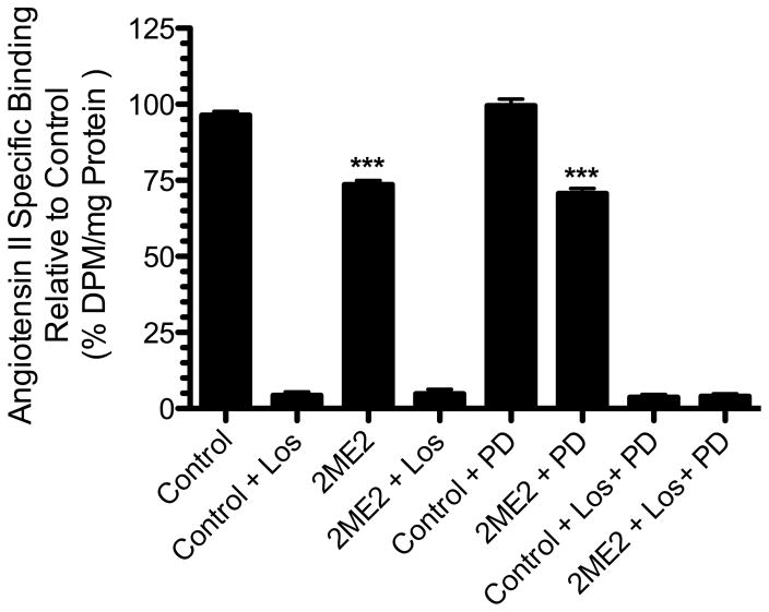
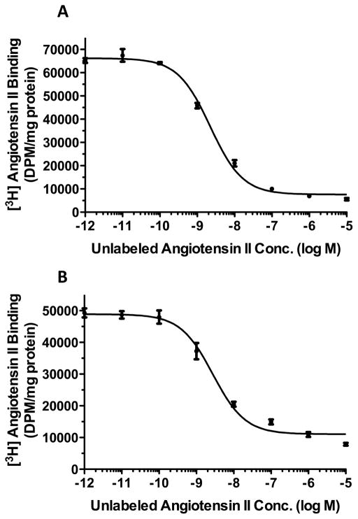
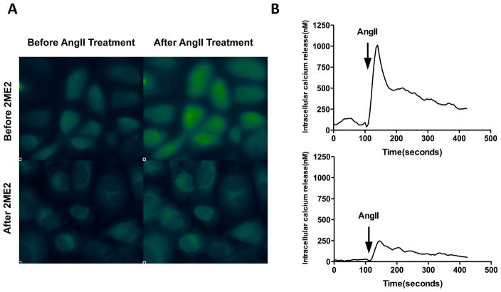
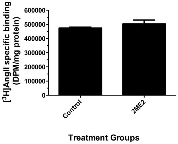
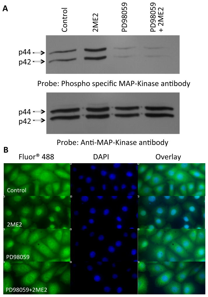
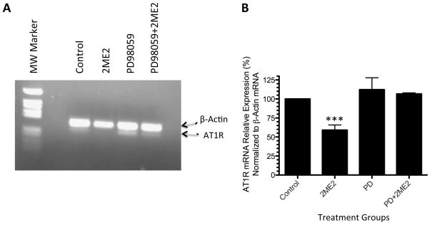
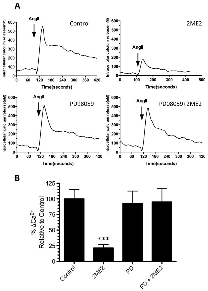
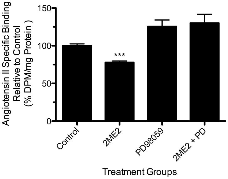
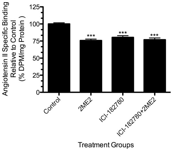
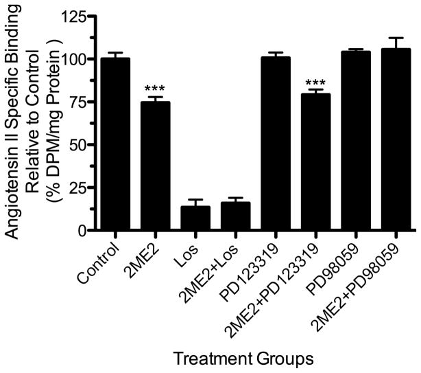
Results: In the presence of 2ME2, cells exhibited significant down-regulation of AngII binding that was both dose and time dependent, independent of estrogen receptors (ERα/ERβ). Down-regulation of AngII binding was AT1R specific, with no change in receptor affinity. Under similar conditions, we observed lower expression of AT1R mRNA, significant inhibition of AngII-mediated increase in intracellular Ca(2+), and increased phosphorylation of ERK1/2. Pretreatment of cells with the MEK inhibitor PD98059 prevented 2ME2-induced ERK1/2 phosphorylation and down-regulation of AT1R expression, which suggests that the observed inhibitory effect is mediated through ERK1/2 signaling intermediates. Similar analyses in stably transfected CHO (Chinese hamster ovary) cell lines with a constitutively active cytomegalovirus promoter showed no change in AT1R expression, which suggests that 2ME2-mediated effects are through transcriptional regulation. The effects of 2ME2 on AT1R down-regulation through ERK1/2 were consistently reproduced in primary rat aortic smooth muscle cells.

Conclusions: Because AT1R has a critical role in the control of CVD, 2ME2-induced changes in receptor expression may provide beneficial effects to the cardiovascular and other systems.


Figures

Figure 1
Figure 1. 2ME2 inhibits AT1R specific binding in WB cells
Radio-ligand binding assay after 24 h 1 μM 2-methoxyestradiol (2ME2) treatment and losartan (Los), or PD123319 (PD) blockade. Cells were exposed to 2ME2 (1 μM) for 24 h and [3H]AngII binding measured in the presence or absence of losartan (an AT1R specific antagonist) or PD123319 (an AT2R specific antagonist). (n=9). Data are expressed as mean ± SEM ***p<0.0001 versus untreated control.
Figure 2
Figure 2. 2ME2 results in AT1R down-regulation in a dose and time dependent manner
(A) 2ME2 mediated AT1R inhibitory effect is dose dependent. Radio-ligand binding assay after variable concentrations (250 – 1000 nM) of 2ME2 treatment for 24 h. (n=9). (B) 2ME2 mediated AT1R inhibitory effect is time dependent. Radio-ligand binding assay after exposure to 1 μM 2ME2 for variable treatment times as indicated. (n=8) Data are expressed as mean ± SEM ***p<0.0001 versus untreated control.
Figure 3
Figure 3. Competition binding studies reveal no change in receptor affinity after 2ME2 treatment
Radio-ligand binding competition studies performed on untreated cells (A) and cells treated with 1 μM 2ME2 for 24 hours (B). Nonlinear least squares regression analysis gave a Kd of 3.321±0.5545 nM for receptors in cells exposed to normal medium and a Kd of 3.568±0.6955 nM for receptors in cells exposed to 2ME2 (n=3).
Figure 4
Figure 4. Down-regulation of AT1R by 2ME2 produces an associated decrease in AngII elicited G-protein coupled response
(A) Fluorescent imaging shows 1 μM 2ME2 exposure for 24 h significantly inhibited AngII mediated increase in intracellular Ca2+, a G-protein coupled response. Transient free Ca2+ was measured with the Ca2+ indicator fura 2-AM using fluorescent imaging microscopy before and after AngII stimulation (10−7 M), with or without 2ME2 treatment. Ca2+ bound fura 2-AM emits green fluorescence. (B) Ca2+ FLIPR assay shows a significant reduction in fluorescent emission after 1 μM 2ME2 exposure for 24 h. Representative tracing of AngII mediated transient increase in intracellular Ca2+ without (upper) and with (lower) 2ME2 treatment. The tracings are representative of 3 separate experiments; an overall reduction of 78.65±16.11%.
Figure 5
Figure 5. 2ME2 treatment does not result in AT1R down-regulation in T3CHO/AT1A linked to CMV promoter
Radio-ligand binding assay performed after 24 h 1 μM 2ME2 treatment. Mean difference between treated and untreated control 5.043±12.94%, p=0.7018 (n=9).
Figure 6
Figure 6. 2ME2 induces phosphorylation of ERK1/2
(A) Total cell lysates were prepared from control, 2ME2 treated, MEK inhibitor PD98059 treated, and 2ME2+PD98059 treated cells and immunoblotted with phospho-specific ERK1/2 antibody (upper). Blot stripped and reprobed with anti-total ERK1/2 antibody to demonstrate equal loading (lower). A representative blot is shown (n=3). (B) ERK1/2 translocates to the nucleus upon treatment with 1 μM 2ME2. Immunofluorescent staining using primary rabbit anti-ERK1/2 IgG followed by secondary anti-rabbit IgG conjugated with Alexa Fluor 488®. Nuclei stained with 4′,6-diamidino-2-phenylindole (DAPI). Treatment groups included are untreated control, 2ME2 treatment, PD98059 treatment, and combined 2ME2 and PD98059 treatment. Images are divided among FITC, DAPI, and overlayed images to demonstrate nuclear co-localization. Representative images are shown of 3 individual experiments.
Figure 7
Figure 7. 2ME2 inhibits AT1R mRNA expression
(A) Representative image of ethidium bromide gel of a dual RT-PCR reaction using AT1R/actin specific primers to determine the effects of 2ME2 on AT1R mRNA in WB cells. Bands detected are at 289 and 206 bp for β-actin and AT1R mRNAs respectively. (B) Quantitation of multiple analyses of AT1R expression were normalized to β-actin and data are expressed as mean ± SEM, (n=3). Densitometric analysis of multiple experiments (N=3) revealed that TA exposure resulted in a 40.99±6.756% (p=0.0037). **p<0.001 compared to untreated control.
Figure 8
Figure 8. Ca2+ FLIPR assay shows MEK inhibitor PD98059 restores intracellular Ca2+ release in 2ME2 treated cells
Increase in intracellular Ca2+ was measured as described in the methods section. (A) Representative tracings of transient increase in intracellular Ca2+ (upper left) in untreated control cells, (upper right) 2ME2 treated cells, (lower left) PD98059 treated cells, and (lower right) PD98059+2ME2 treated cells. (B) Compiled data of 3 experiments and expressed at relative AngII elicited Ca2+ release as compared to untreated control. Mean difference between PD98509+2ME2 and PD treated control 2.209±29.10%, p=0.9431. Data are expressed as mean ± SEM. **p<0.001 compared to control.
Figure 9
Figure 9. MEK inhibition restores radio-ligand binding in 2ME2 treated cells
Radio-ligand binding assay was performed after 24 h 1 μM 2ME2 treated and untreated cells in the presence or absence of PD98059 (20 μM). Mean difference between PD98059+2ME2 and PD98059 control is 4.424±14.60%, p=0.7658 (n=9). Data are expressed as mean ± SEM.
Figure 10
Figure 10. ERα/ERβ antagonism does not restore radio-ligand binding in 2ME2 treated cells
Radio-ligand binding assay was performed after 24 h 1 μM 2ME2 treated and untreated cells in the presence or absence of ICI182780 (50 μM). Mean difference between ICI182780+2ME2 and untreated control is 23.02±3.062%, p<0.001 (n=9). Data are expressed as mean ±SEM.
Figure 11
Figure 11. 2ME2 treatment down-regulates radio-ligand binding in rat aortic smooth muscle cells
Cells were exposed to 2ME2 (1 μM) for 24 h and [3H]AngII binding measured in the presence or absence of losartan (an AT1R specific antagonist) PD123319 (an AT2R specific antagonist), or PD98059 (a specific MEK inhibitor). (n=9). Data are expressed as mean ± SEM ***p<0.0001 versus untreated control.

Similar articles

Cited by

References

    1. Williams GH, Dluhy RG. Disorders of the Adrenal Cortex. 17. New York: McGraw Hill Companies Inc; 2008.
    1. Powers AC. Diabetes Mellitus. 17. New York: McGraw Hill companies Inc; 2008.
    1. Wang CH, Li F, Takahashi N. The renin angiotensin system and the metabolic syndrome. Open Hypertens J. 2010;3:1–13. - PMC - PubMed
    1. Tamura K, Tanaka Y, Tsurumi Y, et al. The role of angiotensin AT1 receptor-associated protein in renin-angiotensin system regulation and function. Curr Hypertens Rep. 2007;9:121–7. - PubMed
    1. Henrion D, Kubis N, Lévy BI. Physiological and pathophysiological functions of the AT(2) subtype receptor of angiotensin II: from large arteries to the microcirculation. Hypertension. 2001;38(5):1150–7. - PubMed

Publication types

MeSH terms

LinkOut - more resources