. 2012 Mar;177(3):638-45.
doi: 10.1016/j.jsb.2012.02.006. Epub 2012 Feb 16.

Crystal structures of the reverse transcriptase-associated ribonuclease H domain of xenotropic murine leukemia-virus related virus

Affiliations

Crystal structures of the reverse transcriptase-associated ribonuclease H domain of xenotropic murine leukemia-virus related virus

Dongwen Zhou et al. J Struct Biol. 2012 Mar.

Abstract

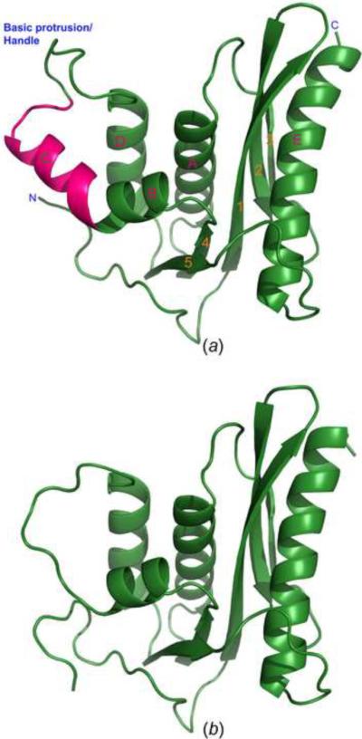
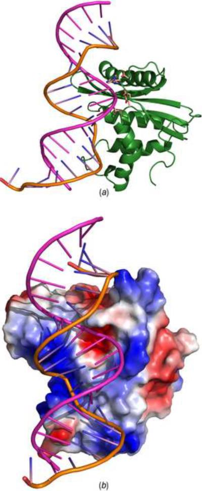
The ribonuclease H (RNase H) domain of retroviral reverse transcriptase (RT) plays a critical role in the life cycle by degrading the RNA strands of DNA/RNA hybrids. In addition, RNase H activity is required to precisely remove the RNA primers from nascent (-) and (+) strand DNA. We report here three crystal structures of the RNase H domain of xenotropic murine leukemia virus-related virus (XMRV) RT, namely (i) the previously identified construct from which helix C was deleted, (ii) the intact domain, and (iii) the intact domain complexed with an active site α-hydroxytropolone inhibitor. Enzymatic assays showed that the intact RNase H domain retained catalytic activity, whereas the variant lacking helix C was only marginally active, corroborating the importance of this helix for enzymatic activity. Modeling of the enzyme-substrate complex elucidated the essential role of helix C in binding a DNA/RNA hybrid and its likely mode of recognition. The crystal structure of the RNase H domain complexed with β-thujaplicinol clearly showed that coordination by two divalent cations mediates recognition of the inhibitor.


Figures

Figure 1
Figure 1
Crystal structures of the full-length XMRV RNase H and its deletion variant ΔC2. (A) Ribbon representation of the overall three-dimensional structure of full-length RNase H. Helix C is highlighted in hot pink, the other parts of the structure in green. Identification of the secondary structure elements (used throughout the manuscript) follows the precedent from related structures. (B) Overall structure of the deletion variant ΔC2 of XMRV RNase H in which helix C was removed. The model is oriented in the same way as in panel A.
Figure 2
Figure 2
Comparisons of the structures of RNase H from different species. (a) Superposition of RNase H from XMRV (green) and E. coli (light pink). (b) Superposition of RNase H from XMRV (green) and HIV-1 (orange). (c) Structure-based sequence alignment of XMRV, human, E. coli, and HIV-1 RNase H enzymes. Catalytic amino acids are highlighted in red, and conserved residues located near the active site and might contribute to catalysis in cyan, whereas other identical residues are highlighted in green. Yellow highlight indicates the sequence corresponding to the helix C and the basic protrusion.
Figure 3
Figure 3
Interactions between the N-terminal loop and the catalytic domain. Arg506 and Asp508 form multiple hydrogen bonds (orange dashed lines) with the residues located in the loop located between helix D and β-strand 5. The 2Fo-Fc electron density map was contoured at 1.0 σ.
Figure 4
Figure 4
(a) Mn2+-dependent RNase H activities of intact HIV-1 and XMRV RT, and a comparison with XMRV variants of the isolated RNase H domain. (b) Inhibition of HIV-1 and XMRV RNase H variants by the active site inhibitor β-thujaplicinol. All enzymes were evaluated in the presence of Mn2+, and IC50 values are the average of triplicate analysis.
Figure 5
Figure 5
Binding of the α-hydroxytropolone inhibitor β-thujaplicinol at the XMRV RNase H active site. Direct contacts between the protein (green), inhibitor (gray), and manganese cations (purple) are shown in blue dashed lines.
Figure 6
Figure 6
A model of the XMRV RNase H-substrate complex. (a) Docking of a 20-mer DNA/RNA hybrid from the experimental structure of its complex with human RNase H (Nowotny et al., 2007) onto XMRV RNase H. The active site residues are shown in sticks. The DNA strand is in orange and the RNA strand is in pink. (b) Surface potential representation of XMRV RNase H complexed to a 20-mer DNA/RNA hybrid (lines). The DNA and RNA strands are colored as in panel A.

References

    1. Adams PD, Grosse-Kunstleve RW, Hung LW, Ioerger TR, McCoy AJ, et al. PHENIX: building new software for automated crystallographic structure determination. Acta Crystallogr. 2002;D58:1948–1954. - PubMed
    1. Boyer PL, Gao HQ, Frank P, Clark PK, Hughes SH. The basic loop of the RNase H domain of MLV RT is important both for RNase H and for polymerase activity. Virol. 2001;282:206–213. - PubMed
    1. Brünger AT. The free R value: a novel statistical quantity for assessing the accuracy of crystal structures. Nature. 1992;355:472–474. - PubMed
    1. Budihas SR, Gorshkova I, Gaidamakov S, Wamiru A, Bona MK, et al. Selective inhibition of HIV-1 reverse transcriptase-associated ribonuclease H activity by hydroxylated tropolones. Nucleic Acids Res. 2005;33:1249–1256. - PMC - PubMed
    1. Cerritelli SM, Crouch RJ. Cloning, expression, and mapping of ribonucleases H of human and mouse related to bacterial RNase HI. Genomics. 1998;53:300–307. - PubMed

Publication types

MeSH terms