Review
. 2012 Oct 1;62(4):2222-31.
doi: 10.1016/j.neuroimage.2012.02.018. Epub 2012 Feb 17.

The Human Connectome Project: a data acquisition perspective

Affiliations
Review

The Human Connectome Project: a data acquisition perspective

D C Van Essen et al. Neuroimage. .

Abstract

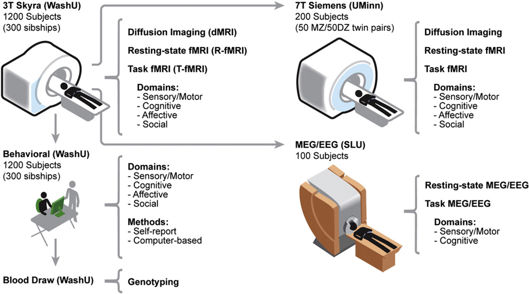
The Human Connectome Project (HCP) is an ambitious 5-year effort to characterize brain connectivity and function and their variability in healthy adults. This review summarizes the data acquisition plans being implemented by a consortium of HCP investigators who will study a population of 1200 subjects (twins and their non-twin siblings) using multiple imaging modalities along with extensive behavioral and genetic data. The imaging modalities will include diffusion imaging (dMRI), resting-state fMRI (R-fMRI), task-evoked fMRI (T-fMRI), T1- and T2-weighted MRI for structural and myelin mapping, plus combined magnetoencephalography and electroencephalography (MEG/EEG). Given the importance of obtaining the best possible data quality, we discuss the efforts underway during the first two years of the grant (Phase I) to refine and optimize many aspects of HCP data acquisition, including a new 7T scanner, a customized 3T scanner, and improved MR pulse sequences.


Figures

Fig. 1
Fig. 1
Schematic summary for acquiring imaging, behavioral, and genetic data using MR and MEG/EEG scanners at three HCP data acquisition sites. Left: Behavioral testing, blood draws for genotyping, and scanning on a 3T Skyra will be carried out on 1200 healthy adults at Washington University (WashU). Center: Major data acquisition modalities are indicated in the center column; for task-fMRI and behavior, major domains are listed. Top right: A subset of 200 subjects will be scanned on a 7T Skyra at the University of Minnesota (UMinn). Bottom right: A subset of 100 subjects will be scanned using magnetoencephalography (MEG) and perhaps electroencephalography (EEG) at St. Louis University (SLU).
Fig. 2
Fig. 2
Relative SNR at the central k space point in diffusion imaging with 150, 100, 70, and 40 mT/m maximum gradients relative to maximum achievable with 300 mT/m when TE is minimized using the available gradient amplitude, calculated for white matter at different b-values ranging from 500 to 10,000.
Fig. 3
Fig. 3
The M-EPI pulse sequence compared with conventional EPI. Top left: EPI pulse sequence generates a single slice image during each readout train, which is repeated for each slice to scan the whole brain. The multiband technique replaces the single slice excitation pulse with multiband (MB) pulses to excite several slices simultaneously, which are then unaliased using array coil sensitivity profiles. As such, far fewer repeats are required to scan the whole brain. Bottom left: Multiplexed-EPI (M-EPI) pulse sequence combines the SIR approach with the MB technique: SIR consecutively excites s slices (s = 3 is shown in the pulse sequence diagram with pulses in red, blue and green) and reads them out in a single echo train, separated in time. Using MB pulses to simultaneously excite m slices instead of exciting each single slice in the SIR approach produces the M-EPI sequence, with a “slice acceleration” of (s × m) leading to (s × m) number of slices collected in a single echo train. Right: Each column shows four (of 60) slices from a whole brain (2 mm isotropic resolution) 3T data set obtained with the M-EPI technique, shown with the (s × m) acceleration factors ranging from 4 to 12. Adapted with permission from Feinberg et al. (2010).

References

    1. Aboitiz F, Scheibel AB, Fisher RS, Zaidel E. Fiber composition of the human corpus callosum. Brain Res. 1992;598:143–153. - PubMed
    1. Achenbach TM, Krukowski RA, Dumenci L, Ivanova MY. Assessment of adult psychopathology: meta-analyses and implications of cross-informant correlations. Psychol. Bull. 2005;131:361–382. - PubMed
    1. Aganj I, Lenglet C, Sapiro G, Yacoub E, Ugurbil K, Harel N. Reconstruction of the orientation distribution function in single- and multiple-shell q-ball imaging within constant solid angle. Magn. Reson. Med. 2010;64:554–566. - PMC - PubMed
    1. Akil H, Martone ME, Van Essen DC. Challenges and opportunities in mining neuroscience data. Science. 2011;331:708–712. - PMC - PubMed
    1. Andersson S, Skare, Ashburner J. How to correct susceptibility distortions in spin-echo echo-planar images: application to diffusion tensor imaging. NeuroImage. 2003;20:870–888. - PubMed

Publication types