. 2012 Jun 15;125(Pt 12):2940-53.
doi: 10.1242/jcs.100834. Epub 2012 Feb 24.

Met receptor tyrosine kinase signals through a cortactin-Gab1 scaffold complex, to mediate invadopodia

Affiliations

Met receptor tyrosine kinase signals through a cortactin-Gab1 scaffold complex, to mediate invadopodia

Charles V Rajadurai et al. J Cell Sci. .

Abstract

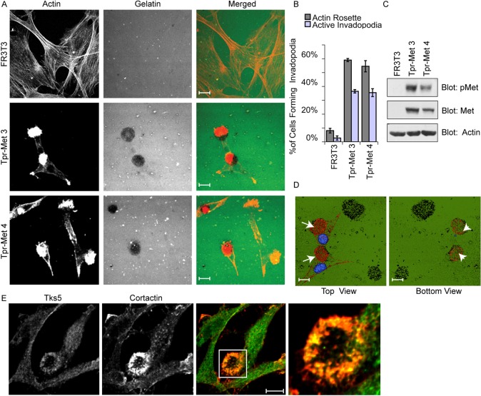
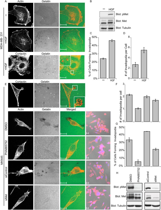
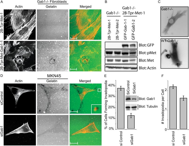
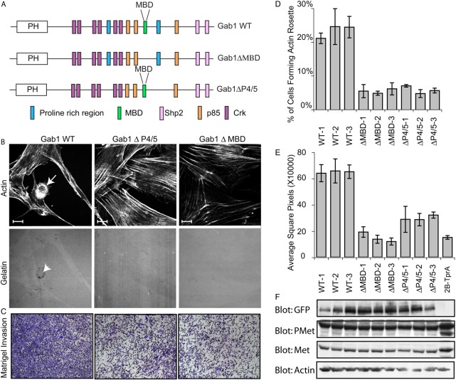
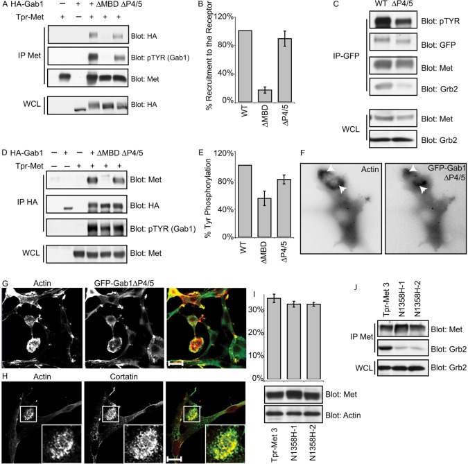
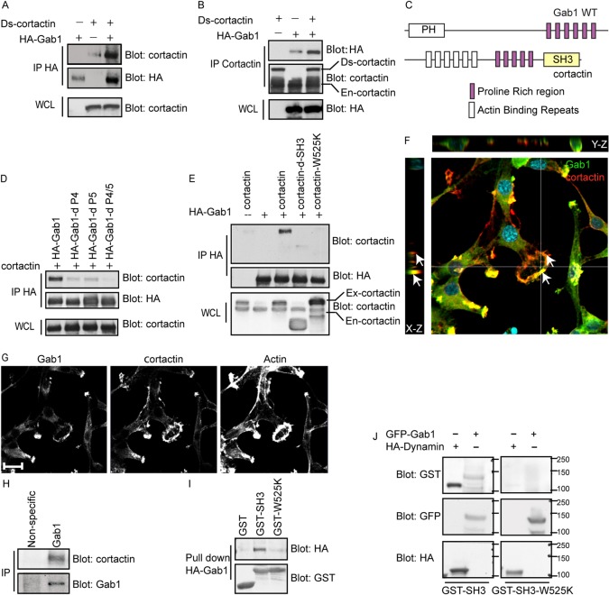
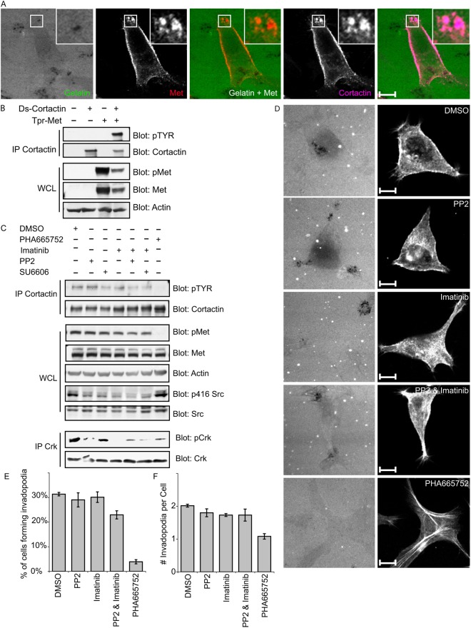
Invasive carcinoma cells form actin-rich matrix-degrading protrusions called invadopodia. These structures resemble podosomes produced by some normal cells and play a crucial role in extracellular matrix remodeling. In cancer, formation of invadopodia is strongly associated with invasive potential. Although deregulated signals from the receptor tyrosine kinase Met (also known as hepatocyte growth factor are linked to cancer metastasis and poor prognosis, its role in invadopodia formation is not known. Here we show that stimulation of breast cancer cells with the ligand for Met, hepatocyte growth factor, promotes invadopodia formation, and in aggressive gastric tumor cells where Met is amplified, invadopodia formation is dependent on Met activity. Using both GRB2-associated-binding protein 1 (Gab1)-null fibroblasts and specific knockdown of Gab1 in tumor cells we show that Met-mediated invadopodia formation and cell invasion requires the scaffold protein Gab1. By a structure-function approach, we demonstrate that two proline-rich motifs (P4/5) within Gab1 are essential for invadopodia formation. We identify the actin regulatory protein, cortactin, as a direct interaction partner for Gab1 and show that a Gab1-cortactin interaction is dependent on the SH3 domain of cortactin and the integrity of the P4/5 region of Gab1. Both cortactin and Gab1 localize to invadopodia rosettes in Met-transformed cells and the specific uncoupling of cortactin from Gab1 abrogates invadopodia biogenesis and cell invasion downstream from the Met receptor tyrosine kinase. Met localizes to invadopodia along with cortactin and promotes phosphorylation of cortactin. These findings provide insights into the molecular mechanisms of invadopodia formation and identify Gab1 as a scaffold protein involved in this process.


Figures

Fig. 1.
Fig. 1.
Tpr-Met-transformed FR3T3 fibroblasts form invadopodia. (A) FR3T3 cells or FR3T3 cells stably overexpressing Tpr-Met (Tpr-Met 3 and Tpr-Met 4) were cultured for 24 hours on glass coverslips coated with Oregon-Green-conjugated gelatin (gelatin matrix). Cells were stained with phalloidin and confocal images were taken at the ventral plane of the cells. Representative images are shown. (B) Quantification of the ability of FR3T3 cells to form actin rosettes or active invadopodia in response to Tpr-Met. Values are the means of three independent experiments. Active invadopodia are defined as actin-rich rosettes overlaying matrix remodeling. (C) SDS-PAGE was performed on cell lysates from FR3T3 cells or FR3T3 cells stably overexpressing Tpr-Met and probed for Met-P (pMet), Met and actin. (D) Confocal Z-sections were collected, deconvolved using IMARIS software and volume rendered to reconstruct the 3D image. View of invadopodia from above (arrows) and below (arrowheads) the gelatin matrix. (E) FR3T3 cells expressing Tpr-Met were plated on glass coverslips coated with unlabeled collagen for 24 hours and immunostained for invadopodia markers Tks5 and cortactin to identify invadopodia rosettes. The boxed region in the third image is shown at higher magnification in the fourth panel. Scale bars: 10 μm.
Fig. 2.
Fig. 2.
Invasive breast cancer cells, MDA-MB-231, and gastric cancer cells, MKN45, form invadopodia in response to Met RTK signaling. (A) MDA-MB-231 cells were cultured on gelatin matrix for 3 hours and stimulated with 0.5 nM HGF for an additional 3 hours. Cells were stained for the invadopodia markers actin (phalloidin) or cortactin. (B) SDS-PAGE was performed on cell lysates of MDA-MB-231 cells stimulated with 0.5 nM HGF and non-stimulated cells. (C,D) The ability of MDA-MB-231 cells to form invadopodia in response to HGF stimulation was quantified. Values are the means of three independent experiments. (E) MKN45 cells were cultured on gelatin matrix in the presence of 0.1 µM Met inhibitor PHA665752 or DMSO for 24 hours. MKN45 cells were treated with 50 nM siRNA targeting Met or control siRNA. Cells were trypsinized 48 hours after treatment and plated on gelatin matrix and cultured for an additional 24 hours. Cells were stained for markers of invadopodia, actin (phalloidin) or cortactin and confocal images were acquired at the ventral plane of the cells. DIC images of cells stained with actin (red) and DAPI (blue) taken at a lower magnification (63×) are shown on the right. Representative images are shown. (F,G) The loss of invadopodia formation in MKN45 cells in response to treatment with Met inhibitor PHA665752 or siRNA-mediated knockdown of Met. (H) SDS-PAGE was performed on cell lysates of MKN45 cells treated with 0.1 µM Met inhibitor or 50 nM siRNA to Met or the respective vehicles (DMSO) or control siRNA and probed for Met-P (pMet), Met and tubulin. Scale bars: 10 μm.
Fig. 3.
Fig. 3.
A multi-substrate docking site on Tpr-Met is required for functional invadopodia formation. (A) FR3T3 cells stably overexpressing various mutants of Tpr-Met with phenylalanine substituted for tyrosine, were cultured on gelatin-matrix-coated coverslips for 24 hours. Cells were stained with phalloidin and confocal images were acquired. Arrows indicate areas of matrix remodeling. Representative images are shown. (B) The ability of Tpr-Met mutants to promote formation of actin rosettes or active invadopodia was quantified. Values are the means of three independent experiments. SDS-PAGE was performed on lysates from cells expressing Tpr-Met mutants and probed for Met and tubulin. Scale bars: 10 μm.
Fig. 4.
Fig. 4.
Gab1 is required for Met-induced invadopodia formation. (A) Gab1 null cells stably overexpressing Tpr-Met (Gab1 null Tpr-Met) and rescued with GFP–Gab1 were cultured on gelatin-matrix-coated coverslips for 24 hours. Cells were stained with phalloidin and confocal images were acquired. (B) Proteins from cell extracts of Gab1 null Tpr-Met cells as well as GFP–Gab1-rescued cells were resolved by SDS-PAGE and immunoblotted for GFP, Met-P (pMet), Met and actin. (C) Gab1 null Tpr-Met cells or cells rescued with GFP–Gab1 were transiently transfected with RFP–actin and subjected to time-lapse video microscopy. One frame from each video is shown. (D) MKN45 cells were treated with 100 nM control siRNA or smartpool siRNA against Gab1 for 48 hours. Cells were trypsinized and plated on gelatin matrix for 24 hours, stained with phalloidin and confocal images were acquired. (E,F) The ability of MKN45 cells to form invadopodia in response to treatment with siRNA against Gab1 or control siRNA was quantified. An immunoblot showing the knockdown of Gab1 in MKN45 cells treated with siRNA against Gab1 but not in control siRNA-treated cells is shown. Scale bars: 10 μm.
Fig. 5.
Fig. 5.
Met-RTK-driven invadopodia biogenesis is dependent on two PxxP motifs in Gab1. (A) Schematic diagram of Gab1, indicating Met binding domain (MBD) and a proline-rich region 4/5 (P4/5) and sites of recruitment for downstream signaling proteins. PH; pleckstrin homology domain. (B) Gab1 null Tpr-Met cells rescued with either WT Gab1, Gab1ΔMBD or Gab1ΔP4/5 were cultured on gelatin matrix for 24 hours, fixed, stained with phalloidin and confocal images acquired. Arrows indicate rosettes and arrowheads indicate matrix remodeling. (C) Cells rescued with either WT Gab1 or the indicated Gab1 mutants were subjected to Boyden chamber invasion assays. Representative images are shown. (D,E) Quantification of invadopodia response (D) and Boyden chamber invasion response (E) is shown for three clones for each mutant and WT Gab1-rescued cells. Values are the means of three independent experiments. (F) SDS-PAGE was performed on lysates from the corresponding rescue cells and probed for GFP, Met-P (pMet), Met and tubulin. Scale bars: 10 μm.
Fig. 6.
Fig. 6.
Gab1ΔP4/5 is recruited to Tpr-Met and localizes to invadopodia but fails to initiate actin rosette formation. (A,D) HEK 293 cells were transiently transfected with the indicated constructs; proteins from lysates were immunoprecipitated with Met 147 antibody (A) or HA antibody (D) and probed as indicated using the Odessey detection system (Li-Cor). (B,E) Densitometric analysis of western blots was performed using Odessey software and tyrosine phosphorylation of Gab1 mutants. Their ability to be recruited to Met is depicted as a percentage of WT Gab1. Values are the means of three independent experiments. (C) Proteins from lysates of Gab-null Tpr-Met cells rescued with GFP–Gab1 (WT) or GFP–Gab1ΔP4/5 were immunoprecipitated with GFP antibody and probed as indicated. (F) FR3T3 Tpr-Met cells were transiently transfected with RFP-actin and GFP-Gab1ΔP4/5 and subjected to time-lapse video microscopy. One frame depicting Gab1ΔP4/5 localization to an actin rosette is shown. (G) FR3T3 Tpr-Met cells were transiently transfected with GFP-Gab1ΔP4/5, plated on gelatin matrix for 24 hours and stained for cortactin and GFP–Gab1. (H) FR3T3 cells stably overexpressing a Tpr-Met mutant that is specifically uncoupled from Grb2 (N1358H) were plated on gelatin matrix for 24 hours and stained for invadopodia markers actin (phalloidin) and cortactin. (I) Ability of Tpr-Met N1358H mutants to induce actin rosette was assessed. Values are the means of two independent clones (FR3T3 Tpr-Met-N1358H-1 and FR3T3 Tpr-Met-N1358H-2). Levels of Tpr-Met were assessed by western blotting. (J) Tpr-Met was immunoprecipitated from lysates prepared from FR3T3 Tpr-Met-N1358H-1 and FR3T3 Tpr-Met-N1358H-2 clones as well as from FR3T3 Tpr-Met 3 cells using Met 147 antibody. SDS-PAGE was performed and proteins immunoblotted as indicated. Scale bars: 10 μm.
Fig. 7.
Fig. 7.
Cortactin interacts with Gab1 through its SH3 domain with proline-rich regions on Gab1. HEK 293 cells were transiently transfected with the indicated constructs; proteins from lysates were immunoprecipitated with HA antibody (A,D,E) or cortactin antibody (B) and immunoblotted as indicated. (C) Schematic diagram depicting a potential interaction of the cortactin SH3 domain and Gab1 proline-rich consensus motifs. (F,G) FR3T3 cells expressing Tpr-Met were transiently transfected with GFP-Gab1 and then plated on gelatin matrix for 24 hours. Cells were stained for GFP, cortactin and phalloidin. (F) X–Z and Y–Z projection of a 40 Z-stack showing the localization of Gab1 and cortactin to membrane protrusions (arrows). (H) Endogenous Gab1 was immunoprecipitated from BT549 cells and probed for cortactin. (I) GST–cortactin SH3 or GST–cortactin SH3-W525K mutant fusion proteins were coupled to GST beads and used to pull down proteins from lysates of HEK 293 cells transiently expressing HA–Gab1. (J) Proteins from HEK 293 cells, transfected with GFP-Gab1 or HA-dynamin, were immunoprecipitated with either HA or GFP antibody. The immune complex was separated by SDS-PAGE and transferred to a nitrocellulose membrane. Nitrocellulose membranes were incubated with fusion proteins of either cortactin GST–SH3 domain or GST–SH3-W525K and immunoblotted as indicated. Scale bars: 10 μm.
Fig. 8.
Fig. 8.
Met colocalizes with cortactin to invadopodia, and cortactin tyrosine phosphorylation is highly dependent on Met kinase activity. (A) MKN45 cells were plated on gelatin matrix for 24 hours and stained for cortactin and Met. The boxed regions are shown enlarged in the insets. (B) HEK 293 cells were transfected with the indicated constructs and the cell extracts were immunoprecipitated with cortactin antibody, 4F11. Immune complexes were separated by SDS-PAGE and probed as indicated. (C) MKN45 cells were treated with 10 µM PP2, 10 µM SU6656, 10 µM Imatinib, 0.1 µM PHA665752 or vehicle (DMSO). Cell extracts were separated by SDS-PAGE and probed as indicated or immunoprecipitated using anti-cortactin (4F11) or anti-Crk antibodies. Immune complexes were separated by SDS-PAGE and probed as indicated. (D) MKN45 cells were plated on gelatin matrix in the presence of 10 µM PP2, 10 µM SU6656, 10 µM Imatinib, 0.1 µM PHA665752 or vehicle (DMSO) for 24 hours and stained with phalloidin. Representative images are shown. (E,F) The ability of MKN45 cells to form invadopodia in the presence of PP2, SU6656, Imatinib or PHA665752 was quantified. Values are the mean of three independent experiments. Scale bars: 10 μm.

Similar articles

Cited by

References

    1. Abella J. V., Vaillancourt R., Frigault M. M., Ponzo M. G., Zuo D., Sangwan V., Larose L., Park M. (2010). The Gab1 scaffold regulates RTK-dependent dorsal ruffle formation through the adaptor Nck. J. Cell Sci. 123, 1306–1319 10.1242/jcs.062570 - DOI - PubMed
    1. Artym V. V., Zhang Y., Seillier–Moiseiwitsch F., Yamada K. M., Mueller S. C. (2006). Dynamic interactions of cortactin and membrane type 1 matrix metalloproteinase at invadopodia: defining the stages of invadopodia formation and function. Cancer Res. 66, 3034–3043 10.1158/0008-5472.CAN-05-2177 - DOI - PubMed
    1. Ayala I., Giacchetti G., Caldieri G., Attanasio F., Mariggiò S., Tetè S., Polishchuk R., Castronovo V., Buccione R. (2009). Faciogenital dysplasia protein Fgd1 regulates invadopodia biogenesis and extracellular matrix degradation and is up-regulated in prostate and breast cancer. Cancer Res. 69, 747–752 10.1158/0008-5472.CAN-08-1980 - DOI - PubMed
    1. Balzer E. M., Whipple R. A., Thompson K., Boggs A. E., Slovic J., Cho E. H., Matrone M. A., Yoneda T., Mueller S. C., Martin S. S. (2010). c-Src differentially regulates the functions of microtentacles and invadopodia. Oncogene 29, 6402–6408 10.1038/onc.2010.360 - DOI - PMC - PubMed
    1. Benvenuti S., Comoglio P. M. (2007). The MET receptor tyrosine kinase in invasion and metastasis. J. Cell. Physiol. 213, 316–325 10.1002/jcp.21183 - DOI - PubMed

Publication types

MeSH terms

Substances