Review
. 2012 Feb 26;15(4):519-27.
doi: 10.1038/nn.3039.

Serotonergic transcriptional networks and potential importance to mental health

Affiliations
Review

Serotonergic transcriptional networks and potential importance to mental health

Evan S Deneris et al. Nat Neurosci. .

Abstract

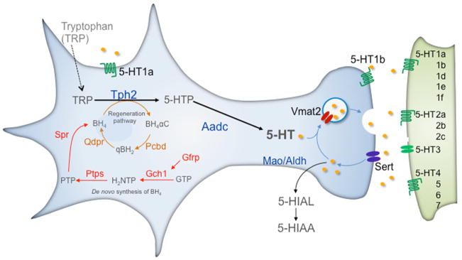
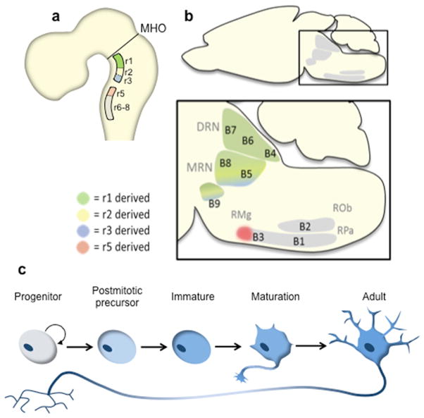
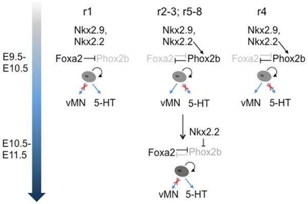
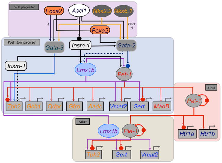
Transcription regulatory networks governing the genesis, maturation and maintenance of vertebrate brain serotonin (5-HT) neurons determine the level of serotonergic gene expression and signaling throughout an animal's lifespan. Recent studies suggest that alterations in these networks can cause behavioral and physiological pathogenesis in mice. Here, we synthesize findings from vertebrate loss-of-function and gain-of-function studies to build a new model of the transcriptional regulatory networks that specify 5-HT neurons during fetal life, integrate them into CNS circuitry in early postnatal life and maintain them in adulthood. We then describe findings from animal and human genetic studies that support possible alterations in the activity of serotonergic regulatory networks in the etiology of mental illness. We conclude with a discussion of the potential utility of our model, as an experimentally well-defined molecular pathway, to predict and interpret the biological effect of genetic variation that may be discovered in the orthologous human network.


Figures

Figure 1
Figure 1. Serotonergic–type neuron identity
5–HT neurons coexpress a gene battery encoding 5–HT synthetic (Tph2, Aadc, Gch1, Gfrp, Ptps, Qdpr), reuptake (Sert), vesicular transport (Vmat2), autoreceptor signaling (Htr1a, Htr1b) and metabolism (Maoa, Maob) proteins. Tetrahydrobiopterin (BH4), an essential cofactor for Tph2 in the synthesis of 5–HTP, is synthesized (red pathway) de novo from guanosine triphosphate (GTP). It is also recycled through a regeneration pathway (brown). Aldehyde dehydrogenase (Aldh) converts 5–Hydroxyindolealdehyde into 5–Hydroxyindoleacetic acid (5–HIAA). Following release, 5–HT modulates 5–HT neuron firing through somatodendritic Htr1a autoreceptors, 5–HT release from the pre–synaptic terminal through Htr1b autoreceptors and stimulates neurotransmission through post–synaptic 5–HT receptors (5–HT1–7). Tph2, Tryptophan hydroxylase 2; Aadc, Aromatic L–amino acid decarboxylase; Sert, Serotonin transporter; Vmat2, vesicular monoamine transporter 2; Mao A,B, Monoamine oxidases A and B; Gch1, GTP cyclohydrolase 1; Gfrp, GTP cyclohydrolase I feedback regulator; Ptps, 6–pyruvoyl–tetrahydropterin synthase; Spr, sepiapterin reductase; Pcbd, pterin–4 alpha–carbinolamine dehydratase; Qdpr, quinoid dihydropteridine reductase. BH4 synthetic intermediates: H2NTP, 7,8–dihydroneopterin triphosphate; Ptp, 6–pyruvoyl–5,6,7,8–tetrahydropterin; BH4αC, tetrahydrobiopterin 4α–carbinolamine; qBH2, Quinoid–dihydrobiopterin.
Figure 2
Figure 2. Neuroanatomical features of 5–HT neuron development
a) Schematic sagittal view of the developing rodent brain. All rodent 5–HT neurons are born caudal to the mid–hindbrain organizer (MHO) in two longitudinal domains, rostral and caudal, on either side of the floor plate in the ventral rhombencephalon (hindbrain). b) Schematic sagittal view of the adult midbrain, pons and medulla depicting the location of 5–HT neurons clusters (raphe nuclei). Intersectional/substractive fate mapping has shown that all 5–HT neurons in the mature dorsal raphe nucleus (DRN, B4, B6 and B7 groups) are born in r1 (green highlight in a and b). 5–HT neurons in the median raphe nucleus (MRN, B8 and B5 groups) and laterally extending supraleminscal 5–HT neurons, designated the B9 cluster, are derived from serotonergic progenitors in r1, r2 and r3 (green, yellow, blue highlight in a and b). 5–HT neurons born in r5–r8 migrate to form the raphe pallidus (RPa, B1), raphe obscurus (ROb, B2), raphe magnus (RMg, B3) and cell bodies in the ventrolateral medulla (B3). 5–HT neurons in the rostral portion of B3 are born in r5 (red highlight). c) Stages of 5–HT neuron development. Postmitotic 5–HT neuron precursors do not yet possess overt serotonergic identity. Induction of the serotonergic–type gene battery generates immature 5–HT neurons. These newborn neurons are integrated with CNS circuitry during a prolonged maturation stage that produces adult 5–HT neurons.
Figure 3
Figure 3. Serotonergic progenitor specification
In rhombomere 1 (r1), Foxa2 continually suppresses Phox2b expression to block production of visceral motor neuron (vMN) progenitors and promotes production of 5–HT progenitors. In r2–3, r5–r8, Nkx2.2 initially induces Phox2b, which in turn represses Foxa2. This subcircuit establishes a motoneuron fate of ventral progenitors in r2–r3 and r5–r8. These progenitors produce motor neurons from E9.5 to about E10.5. Starting at about E10.5 a reciprocal temporal cross–repression is initiated whereby Foxa2 now shuts off Phox2b expression. Foxa2 and Nkx2.2 mediated repression of Phox2b switches the fate of ventral progenitors so that a wave of serotonergic neurogenesis follows motor neurogenesis. In r4, Phox2b continually represses Foxa2 thus suppressing the vMN to 5–HT fate switch and prolonging the production of vMNs.
Figure 4
Figure 4. Serotonergic transcription regulatory network
Stage–specific subcircuits in the network are depicted in colored rectangles with enclosed transcriptional factor gene symbols and arrows or blocked lines leading to other network constituents. Foxa2 is required for Gata3 expression only in r1. In mouse Nkx6.1 and Nkx6.2 do not appear to be required for 5–HT neuron generation but in r4 they suppress 5–HT fate. Nkx6.1 has been shown to be required for Gata2 and Pet–1 expression and production of 5–HT neurons only in chick r1. Chromatin immunoprecipitation experiments indicate direct Gata2 binding (solid blue circle), in vivo, to the Pet–1 upstream regulatory region where conserved Gata2 binding sites are located. Pet–1 induces Htr1a and Htr1b to initiate formation of the two 5–HT autoreceptor pathways in maturing 5–HT neurons (red highlighted box, E14.5). ChIP indicates that Pet–1 binds (solid red circles), in vivo, to the Tph2, Sert, and Pet–1 upstream regions where conserved Pet–1 binding are located. In adult 5–HT neurons (beige highlighted box) Lmx1b and Pet–1 maintain Tph2 and Sert. Lmx1b also maintains Vmat2 expression. Pet–1 expression in adult 5–HT neurons is stabilized through direct positive autoregulation (curved red arrow). Dashed arrows indicates that Gata2 is likely required for Lmx1b expression but this has not been formally demonstrated and Insm–1 may also directly regulate Tph2 but this has not been shown with ChIP.

Similar articles

Cited by

References

    1. Gaddum JH. Ciba Foundation Symposium on Hypertension. Little, Brown and Co; 1954. Drugs antagonistic to 5-hydroxytryptamine; pp. 75–77.
    1. Woolley DW, Shaw E. A Biochemical and Pharmacological Suggestion About Certain Mental Disorders. Proceedings of the National Academy of Sciences of the United States of America. 1954;40:228–231. - PMC - PubMed
    1. Muller CP, Jacobs BL, editors. Handbook of the Behavioral Neurobiology of Serotonin. Academic Press; 2010.
    1. Ishimura K, et al. Quantitative analysis of the distribution of serotonin-immunoreactive cell bodies in the mouse brain. Neurosci Lett. 1988;91:265–270. - PubMed
    1. Baker KG, et al. Cytoarchitecture of serotonin-synthesizing neurons in the pontine tegmentum of the human brain. Synapse. 1991;7:301–320. - PubMed

Publication types