. 2012 Mar;135(Pt 3):736-50.
doi: 10.1093/brain/awr361.

Frontotemporal dementia with the C9ORF72 hexanucleotide repeat expansion: clinical, neuroanatomical and neuropathological features

Affiliations

Frontotemporal dementia with the C9ORF72 hexanucleotide repeat expansion: clinical, neuroanatomical and neuropathological features

Colin J Mahoney et al. Brain. 2012 Mar.

Abstract

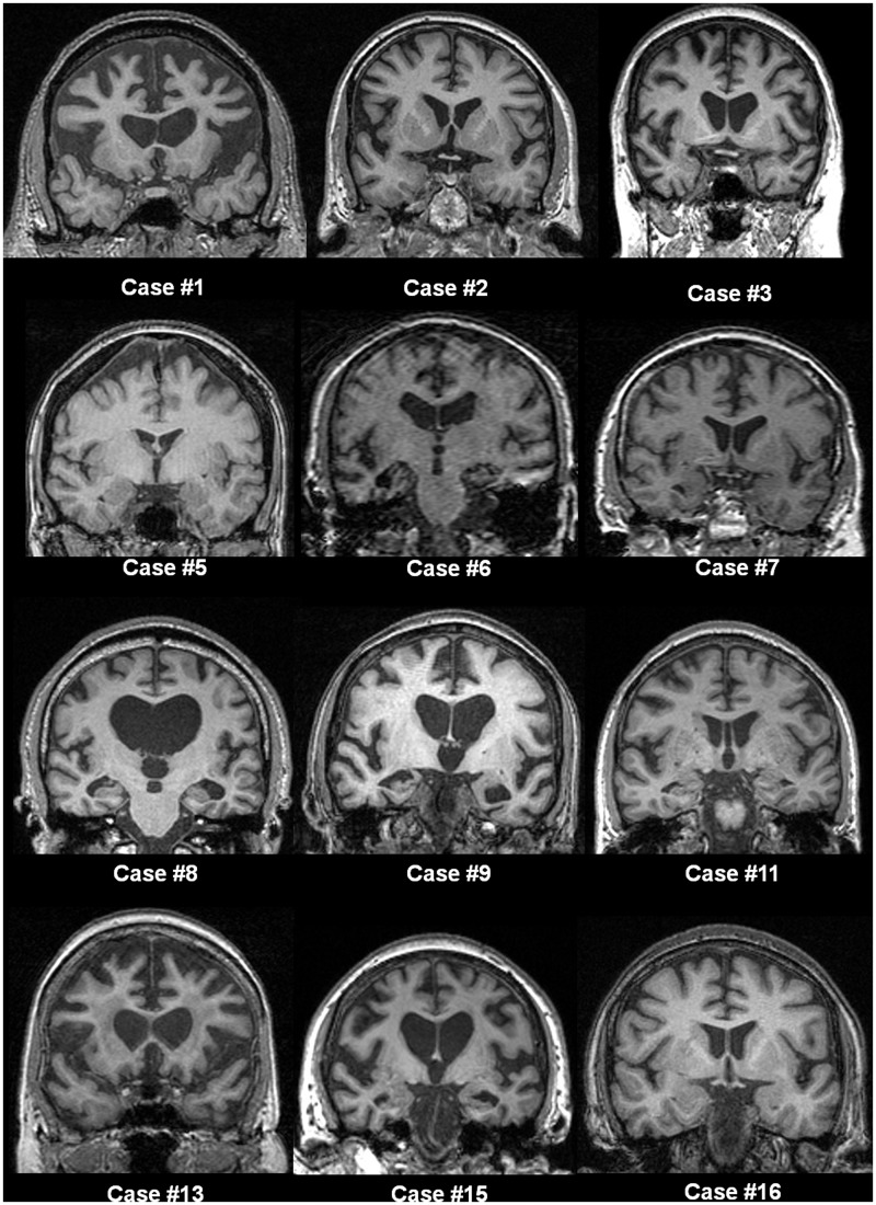
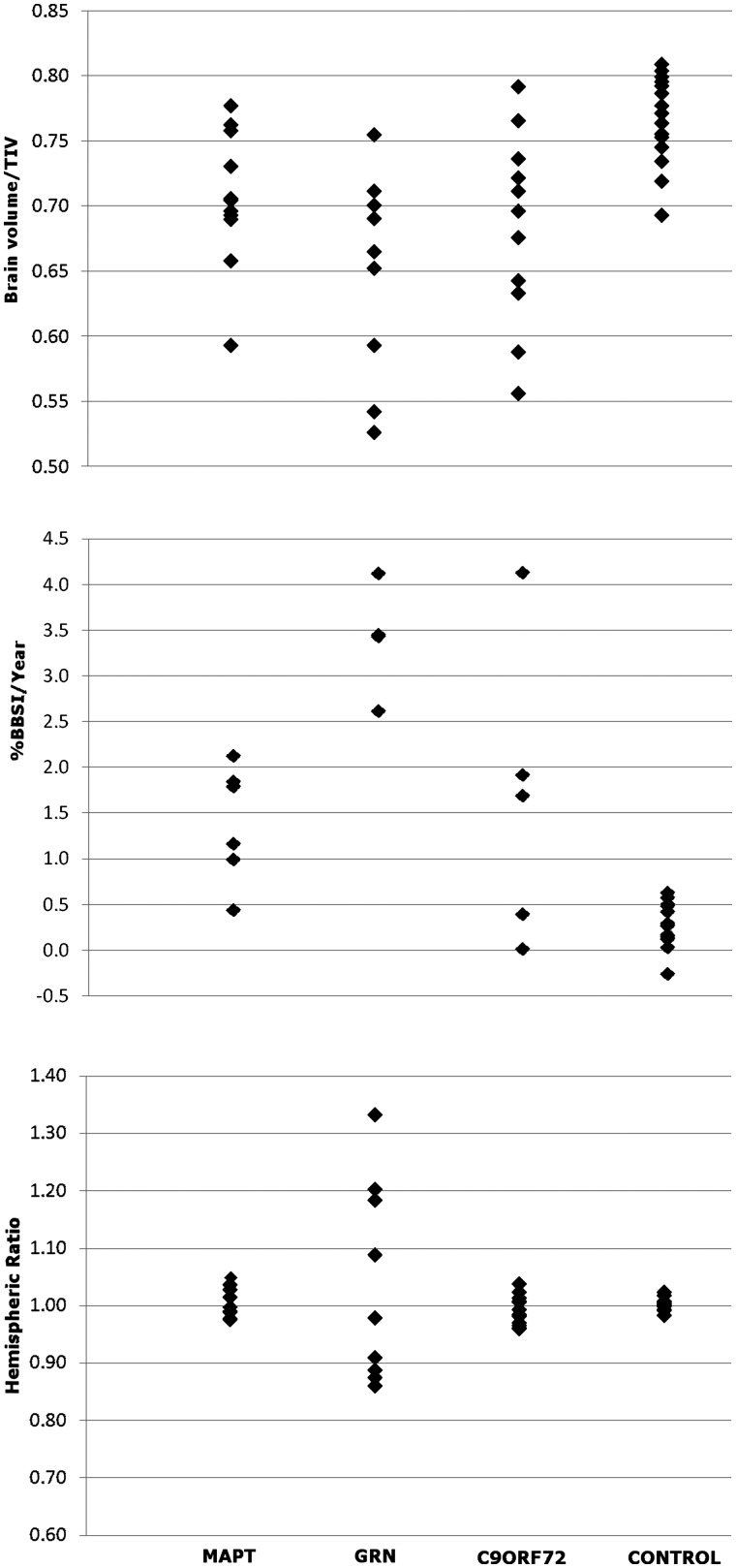
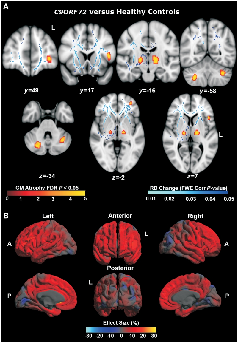
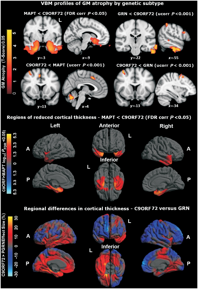
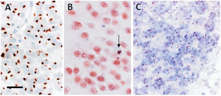
An expanded hexanucleotide repeat in the C9ORF72 gene has recently been identified as a major cause of familial frontotemporal lobar degeneration and motor neuron disease, including cases previously identified as linked to chromosome 9. Here we present a detailed retrospective clinical, neuroimaging and histopathological analysis of a C9ORF72 mutation case series in relation to other forms of genetically determined frontotemporal lobar degeneration ascertained at a specialist centre. Eighteen probands (19 cases in total) were identified, representing 35% of frontotemporal lobar degeneration cases with identified mutations, 36% of cases with clinical evidence of motor neuron disease and 7% of the entire cohort. Thirty-three per cent of these C9ORF72 cases had no identified relevant family history. Families showed wide variation in clinical onset (43-68 years) and duration (1.7-22 years). The most common presenting syndrome (comprising a half of cases) was behavioural variant frontotemporal dementia, however, there was substantial clinical heterogeneity across the C9ORF72 mutation cohort. Sixty per cent of cases developed clinical features consistent with motor neuron disease during the period of follow-up. Anxiety and agitation and memory impairment were prominent features (between a half to two-thirds of cases), and dominant parietal dysfunction was also frequent. Affected individuals showed variable magnetic resonance imaging findings; however, relative to healthy controls, the group as a whole showed extensive thinning of frontal, temporal and parietal cortices, subcortical grey matter atrophy including thalamus and cerebellum and involvement of long intrahemispheric, commissural and corticospinal tracts. The neuroimaging profile of the C9ORF72 expansion was significantly more symmetrical than progranulin mutations with significantly less temporal lobe involvement than microtubule-associated protein tau mutations. Neuropathological examination in six cases with C9ORF72 mutation from the frontotemporal lobar degeneration series identified histomorphological features consistent with either type A or B TAR DNA-binding protein-43 deposition; however, p62-positive (in excess of TAR DNA-binding protein-43 positive) neuronal cytoplasmic inclusions in hippocampus and cerebellum were a consistent feature of these cases, in contrast to the similar frequency of p62 and TAR DNA-binding protein-43 deposition in 53 control cases with frontotemporal lobar degeneration-TAR DNA-binding protein. These findings corroborate the clinical importance of the C9ORF72 mutation in frontotemporal lobar degeneration, delineate phenotypic and neuropathological features that could help to guide genetic testing, and suggest hypotheses for elucidating the neurobiology of a culprit subcortical network.


Figures

Figure 1
Figure 1
Examples of electrophoresed products of repeat-primed polymerase chain reaction used to detect expansions in C9ORF72. Each panel was scaled to 800 fluorescent units on the y-axis and between 300–650 bp on the x-axis. (A) Data from a FTLD patient sample without an expansion and (B) data from a patient with FTLD with 2 and >60 repeats.
Figure 2
Figure 2
FTLD DNA cohort by modified Goldman score. Each bar displays % of cohort by assigned Goldman score and mutation status.
Figure 3
Figure 3
Representative pedigrees from the Dementia Research Centre (DRC) cohort indicating inheritance, predominant clinical syndrome and, where known, age at onset (RIP indicates only age at death known). bvFTD = behavioural variant FTD.
Figure 4
Figure 4
Prevalence of symptoms and neurological signs (expressed as proportion of disease cohort) observed in C9ORF72 cases. Data for first and final assessments are coded separately.
Figure 5
Figure 5
Representative T1-weighted coronal magnetic resonance brain images for cases with C9ORF72 mutation. Numbering of cases is as per Table 2. Patterns of atrophy were highly variable across the group, and no clear consistent profile of atrophy was evident visually. The distribution of atrophy was not prominently asymmetric between the cerebral hemispheres in most cases. Several cases had predominantly frontal lobe atrophy (Cases 1, 2 and 13), while temporal lobe atrophy was prominent in other cases (Cases 6 and 9). Other cases showed more generalized involvement with more prominent central atrophy (Cases 8 and 15) or minimal cross-sectional atrophy to visual inspection (Case 16).
Figure 6
Figure 6
Individual brain volumes corrected for total intracranial volume (TIV) by group (top). Annualized percentage change in brain boundary shift integral (BBSI) by group (middle). Hemispheric asymmetry expressed as the ratio of left/right hemisphere volumes by subject and group (bottom).
Figure 7
Figure 7
(A) Profiles of grey matter (GM) atrophy (n = 11) and increased radial diffusivity (RD) maps (n = 3) in C9ORF72 cases versus healthy controls. MNI co-ordinates are displayed. (B) Mean difference in cortical thickness expressed as a percentage of the mean thickness in C9ORF72 cases (n = 11) versus controls. A = anterior; FDR = false discovery rate; FWE = family-wise error; L = left; P = posterior.
Figure 8
Figure 8
Top: Voxel-based morphometry (VBM) profiles of grey matter (GM) atrophy by genetic subtype; P < 0.001. MNI co-ordinates are displayed. Middle: regional reduction of cortical thickness in MAPT mutation carriers compared with C9ORF72 mutation carriers. Colour scale for statistical difference represents FDR-corrected P < 0.05. Bottom: regional variation of cortical thickness in GRN mutation carriers compared with C9ORF72 mutation carriers. Colour bar for per cent difference represents magnitude of cortical thickness group difference expressed as a percentage of the mean group thickness. Red and yellow (positive values) represent lower cortical thickness in the GRN group, whereas dark to light blue (negative values) represent lower cortical thickness in the C9ORF72 group. A = anterior; FDR = false discovery rate; L = left; P = posterior; Ucorr = uncorrected.
Figure 9
Figure 9
P62 and TDP-43 immunohistochemistry in FTLD-TDP with the C9ORF72 hexanucleotide repeat expansion. Frequent p62-positive (A) and sparse TDP-43-positive (B, arrow) neuronal cytoplasmic inclusions were present in the granule cells of the dentate gyrus in a case with the C9ORF72 repeat expansion. Numerous p62-positive neuronal cytoplasmic inclusions were also present in the cerebellar granule cell (C), which were negative for TDP-43. Scale bars: A = 50 µm; B = 30 µm; C = 80 µm.

References

    1. Al-Sarraj S, King A, Troakes C, Smith B, Maekawa S, Bodi I, et al. p62 positive, TDP-43 negative, neuronal cytoplasmic and intranuclear inclusions in the cerebellum and hippocampus define the pathology of C9orf72-linked FTLD and MND/ALS [Internet] Acta Neuropathologica. 2011 Available from: http://www.springerlink.com/index/10.1007/s00401-011-0911-2 (23 November 2011, date last accessed) - DOI - PubMed
    1. Andreasen NC, O'Leary DS, Cizadlo T, Arndt S, Rezai K, Ponto LL, et al. Schizophrenia and cognitive dysmetria: a positron-emission tomography study of dysfunctional prefrontal-thalamic-cerebellar circuitry. Proc Natl Acad Sci USA. 1996;93:9985–90. - PMC - PubMed
    1. Beck J, King A, Scahill R, Warren JD, Fox NC, Rossor MN, et al. A distinct clinical, neuropsychological and radiological phenotype is associated with progranulin gene mutations in a large UK series. Brain. 2008;131:706–20. - PMC - PubMed
    1. Boeve BF, Lang AE, Litvan I. Corticobasal degeneration and its relationship to progressive supranuclear palsy and frontotemporal dementia. Ann Neurol. 2003;54(Suppl 5):S15–S19. - PubMed
    1. Boxer AL, Mackenzie IR, Boeve BF, Baker M, Seeley WW, Crook R, et al. Clinical, neuroimaging and neuropathological features of a new chromosome 9p-linked FTD-ALS family. J Neurol Neurosurg Psychiatr. 2011;82:196–203. - PMC - PubMed

Publication types

MeSH terms