. 2012 Mar;135(Pt 3):886-99.
doi: 10.1093/brain/aws012.

NADPH oxidase expression in active multiple sclerosis lesions in relation to oxidative tissue damage and mitochondrial injury

Affiliations

NADPH oxidase expression in active multiple sclerosis lesions in relation to oxidative tissue damage and mitochondrial injury

Marie T Fischer et al. Brain. 2012 Mar.

Abstract

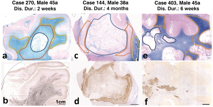
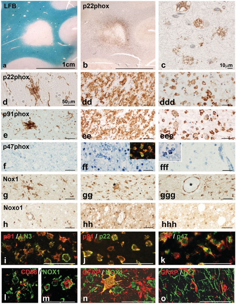
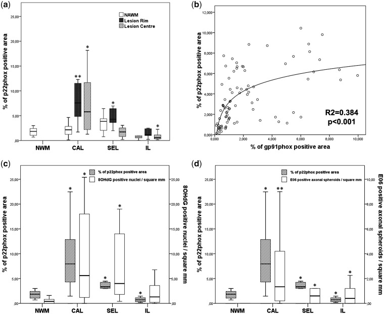
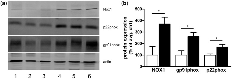
Multiple sclerosis is a chronic inflammatory disease of the central nervous system, associated with demyelination and neurodegeneration. The mechanisms of tissue injury are poorly understood, but recent data suggest that mitochondrial injury may play an important role in this process. Mitochondrial injury can be triggered by reactive oxygen and nitric oxide species, and we recently provided evidence for oxidative damage of oligodendrocytes and dystrophic axons in early stages of active multiple sclerosis lesions. In this study, we identified potential sources of reactive oxygen and nitrogen species through gene expression in carefully staged and dissected lesion areas and by immunohistochemical analysis of protein expression. Genome-wide microarrays confirmed mitochondrial injury in active multiple sclerosis lesions, which may serve as an important source of reactive oxygen species. In addition, we found differences in the gene expression levels of various nicotinamide adenine dinucleotide phosphate oxidase subunits between initial multiple sclerosis lesions and control white matter. These results were confirmed at the protein level by means of immunohistochemistry, showing upregulation of the subunits gp91phox, p22phox, p47phox, nicotinamide adenine dinucleotide phosphate oxidase 1 and nicotinamide adenine dinucleotide phosphate oxidase organizer 1 in activated microglia in classical active as well as slowly expanding lesions. The subunits gp91phox and p22phox were constitutively expressed in microglia and were upregulated in the initial lesion. In contrast, p47phox, nicotinamide adenine dinucleotide phosphate oxidase 1 and nicotinamide adenine dinucleotide phosphate oxidase organizer 1 expression were more restricted to the zone of initial damage or to lesions from patients with acute or early relapsing/remitting multiple sclerosis. Double labelling showed co-expression of the nicotinamide adenine dinucleotide phosphate oxidase subunits in activated microglia and infiltrated macrophages, suggesting the assembly of functional complexes. Our data suggest that the inflammation-associated oxidative burst in activated microglia and macrophages plays an important role in demyelination and free radical-mediated tissue injury in the pathogenesis of multiple sclerosis.


Figures

Figure 1
Figure 1
Acute multiple sclerosis lesions used for gene expression analysis; the structure of the lesions is shown in sections stained with Luxol fast blue (myelin; a, c and e); the lower panel of figures shows the same lesions, stained for p22phox expression in activated macrophages and microglia. In the first patient (a and b), the active lesion (black outline) is surrounded by a broad area of microglia activation with p22phox expression and myelin pallor (red outline; initial lesion), which makes it difficult to see the lesion margin in the staining for macrophages and microglia. In the normal-appearing white matter (yellow outline), myelin density is normal, but there is still moderate microglia activation. In the second patient (c and d), the demyelinated lesion core (black outline) shows concentric rings of preserved myelin. This is surrounded by the initial lesion area with extensive immunoreactivity for p22phox (red outline). The normal-appearing white matter shows normal myelin density and low expression of macrophage antigens (yellow outline). In the third patient (e and f) a dense infiltrate of macrophages with p22phox expression is seen in the area of demyelination (active plaque). In the surrounding white matter, there is little expression of macrophage/microglia antigens. Areas of normal white matter used for gene expression analysis are shown by the yellow outline. Gene expression for proteins involved in oxidative damage and for mitochondrial proteins has been analysed separately in the indicated lesion areas. Dis. Dur = disease duration.
Figure 2
Figure 2
(a–c) Active lesion in a patient with primary progressive multiple sclerosis. (a) Luxol fast blue (LFB) myelin staining shows a demyelinated lesion with defined borders. (b) In the adjacent section stained for p22phox intense expression is seen at the active lesion edge, spanning into the adjacent normal appearing white matter (initial lesion area). (c) In the inactive centre of the lesion p22phox is weakly expressed in some macrophages. [d–h(hh)] These images show the expression of oxidative burst associated molecules in the normal appearing white matter (left column), in the zone of initial tissue injury (centre column) and in the demyelinated zone (right column). Most pronounced expression of all proteins is seen in the initial lesion area (centre panel), while expression for p22phox and gp91phox is much weaker in lipid containing macrophages in the lesion centre (right panel). In the normal appearing white matter microglia nodules can be seen, which are intensely stained for p22phox, gp91phox and NOX1. P22phox, gp91phox and p47phox are only expressed in macrophages and microglia (see below), while Nox1 shows a broader expression also in astrocytes (asterisk in gg) and endothelial cells (asterisk labels the vessel with endothelial staining); the expression of Noxo1 is even broader compared with that of Nox1. P47phox staining is absent in the normal appearing white matter (g), while intense expression is seen in macrophages and small microglia like cells; the insert shows expression of p47phox (red) in macrophages stained with LN3 (green). In the lesion centre, only weak reactivity for p47phox is seen, mainly in perivascular macrophages (at higher magnification in the inset). (l–o) Confocal laser microscope images of double staining with different Nox markers and CNS cell-specific markers; the staining combinations are indicated on the figures. These data show co-localization of different Nox components within the same macrophages or microglia cells in the multiple sclerosis lesions. Nox 1 is also expressed in GFAP-positive astrocytes (n) in the absence of p22phox (o). Red and green staining depicts the individual antigens as indicated in the figure; yellow staining represents double staining.
Figure 3
Figure 3
(a) Quantitative analysis of p22phox expression in different types of multiple sclerosis lesions. Compared with control white matter, there is a significantly higher expression (P < 0.01) in classical active (CAL) and slowly expanding multiple sclerosis lesions (SEL), and in the normal-appearing white matter (NWM) of slowly expanding lesions; in the lesions p22phox-positive microglia are mainly seen in the active lesion edge (initial lesions, IL) and less in the inactive lesion centre. Furthermore, we found a significant decrease of p22 expressing microglia in the centre of inactive lesions. (b) Correlation between p22 and gp91 expression in different multiple sclerosis cases and lesions. The same areas of normal-appearing white matter and lesions were scanned for p22phox and gp91phox expression and regression was analysed as described in the ‘Material and methods’ section. (c) Comparison between p22phox expression, determined by densitometry and the number of nuclei with oxidized DNA (8OHdG immunoreactivity) within multiple sclerosis lesions. (d) Comparison between p22phox expression and the number of dystrophic axons, immunoreactive for oxidized phospholipids (E06); p22phox and gp91phox immunoreactivity was determined by densitometry; nuclei with oxidized DNA and dystrophic axons, positive for E06 were counted manually (Haider et al., 2011). *P<0.05; **P<0.01.
Figure 4
Figure 4
(a) Western blot of three control samples (Lanes 1–3) and three multiple sclerosis samples (Lanes 4–6) demonstrating enhanced protein expression of NOX1, p22phox and gp91phox in active demyelinating multiple sclerosis lesions compared with white matter from non-neurological controls. (b) Quantitative densitometry of the blots reveals significantly increased expression levels for NOX1, gp91phox and p22phox in multiple sclerosis lesions compared with control white matter. *P.

Comment in

References

    1. Bagasra O, Michaels FH, Zheng YM, Bobroski LE, Spitsin SV, Fu ZF, et al. Activation of the inducible form of nitric oxide synthase in the brains of patients with multiple sclerosis. Proc Natl Acad Sci USA. 1995;92:12041–5. - PMC - PubMed
    1. Barnett MH, Prineas JW. Relapsing and remitting multiple sclerosis: pathology of the newly forming lesion. Ann Neurol. 2004;55:458–68. - PubMed
    1. Bauer J, Elger CE, Hans VH, Schramm J, Urbach H, Lassmann H, et al. Astrocytes are a specific immunological target in Rasmussen's encephalitis. Ann Neurol. 2007;62:67–80. - PubMed
    1. Becanovic K, Jagodic M, Sheng JR, Dahlman I, Aboul Enein F, Wallstrom E, et al. Advanced intercross line mapping of Eae5 reveals Ncf-1 and CLDN4 as candidate genes for experimental autoimmune encephalomyelitis. J Immunol. 2006;176:6055–64. - PubMed
    1. Bedard K, Krause KH. The NOX family of ROS-generating NADPH oxidases: physiology and Pathophysiology. Physiol Rev. 2007;87:245–313. - PubMed

Publication types

MeSH terms