

Low-grade malignant myoepithelioma arising in a pleomorphic adenoma: a rare case
- PMID: 22366836
- PMCID: PMC6086638
- DOI: 10.5144/0256-4947.2012.209
Low-grade malignant myoepithelioma arising in a pleomorphic adenoma: a rare case
Abstract
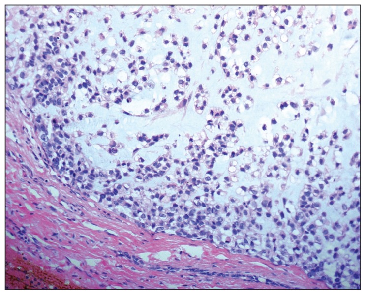
Malignant myoepithelioma is a very rare salivary gland tumor that can arise de novo or within a pre-existing pleomorphic adenoma. We report a case of malignant myoepithelioma most probably arising in a pre-existing pleomorphic adenoma of the left parotid gland. The patient was a 60-year-old man who presented with a multinodular mass lesion over left side of the face and neck. He had undergone removal of a pleomorphic adenoma of the left parotid gland twice (8 and 22 years ago). Histological examination showed locally concentrated highly invasive myoepithelial cells with bland-looking morphology and no evidence of mitosis or necrosis. Immunohistochemistry confirmed the myoepithelial differentiation (S- 100+, SMA+) and a low Ki-67 labeling index (<5%).
Figures


Similar articles
-
A case report of metastasizing myoepithelial carcinoma of the parotid gland arising in a recurrent pleomorphic adenoma.Auris Nasus Larynx. 2009 Feb;36(1):123-6. doi: 10.1016/j.anl.2008.05.014. Epub 2008 Jul 22. Auris Nasus Larynx. 2009. PMID: 18650039
-
Epithelial myoepithelial carcinoma of the parotid gland with malignant ductal and myoepithelial components arising in a pleomorphic adenoma: a case report with cytologic, histologic and immunohistochemical correlation.Acta Cytol. 2007 Sep-Oct;51(5):807-13. doi: 10.1159/000325847. Acta Cytol. 2007. PMID: 17910353
-
Malignant myoepithelial carcinoma (myoepithelioma) arising in a pleomorphic adenoma of the parotid gland. An immunohistochemical study and review of the literature.Oral Surg Oral Med Oral Pathol. 1988 Jul;66(1):65-70. doi: 10.1016/0030-4220(88)90069-2. Oral Surg Oral Med Oral Pathol. 1988. PMID: 2457198 Review.
-
Myoepithelial carcinoma (malignant myoepithelioma) of the parotid gland arising in a pleomorphic adenoma.J Clin Pathol. 1998 Jul;51(7):552-6. doi: 10.1136/jcp.51.7.552. J Clin Pathol. 1998. PMID: 9797738 Free PMC article.
-
Malignant myoepithelioma of salivary glands: clinicopathological features of ten cases.Virchows Arch A Pathol Anat Histopathol. 1993;423(5):389-96. doi: 10.1007/BF01607152. Virchows Arch A Pathol Anat Histopathol. 1993. PMID: 8116228 Review.
References
-
- Dardick I. A role for electron microscopy in salivary gland neoplasms. Ultrastruct Pathol. 1985;9:151–161. - PubMed
-
- Seifert G, Sobin LH. The world health organization’s histological classification of salivary tumors. Cancer. 1992;70:379–385. - PubMed
-
- Ellis GL, Auclair PL. Tumors of the salivary glands. Washington, DC: AFIP; 1996.
-
- Nagao T, Sugano I, Ishida Y. Salivary gland malignant myoepithelioma: A clinicopathological and immunohistochemical study of ten cases. Cancer. 1998;83:1292–1299. - PubMed
-
- Savera AT, Sloman A, Huvos AG. Myoepithelial carcinoma of the salivary glands: A clinicopathologic study of 25 cases. Am J Surg Path. 2000;24:761–774. - PubMed
MeSH terms
LinkOut - more resources
Full Text Sources