

Enhanced activation of human dendritic cells by silencing SOCS1 and activating TLRs simultaneously
- PMID: 22366886
- PMCID: PMC11028872
- DOI: 10.1007/s00262-012-1218-4
Enhanced activation of human dendritic cells by silencing SOCS1 and activating TLRs simultaneously
Abstract
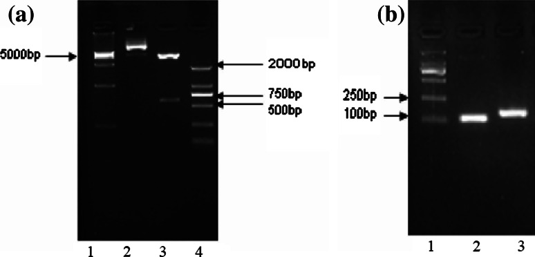
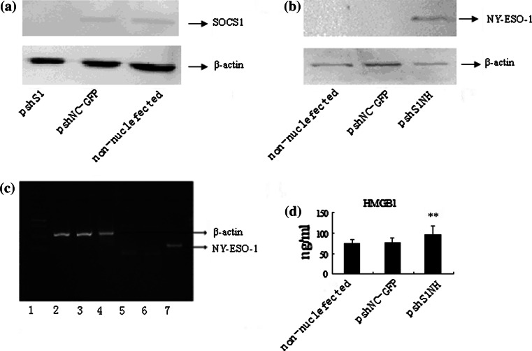
There was established evidence that silencing the attenuator and activating the TLRs could activate the dendritic cells in synergic effects. In this study, we constructed a plasmid, namely pshS1NH, which encodes SOCS1-shRNA, NY-ESO-1-MAGE3 (HLA-A2*0201) fusion antigen and secretory HMGB1, an agent used to modify dendritic cells (DCs), aiming to generate potent DC vaccine against tumors. The SOCS1-shRNA could efficiently downregulate the expression of SOCS1, as indicated by real-time RT-PCR and Western blot. The fusion antigen was detected in the pshS1NH-DCs by PCR and Western blot. Simultaneously, HMGB1 level in the pshS1NH-DCs culture media was significantly higher than that in the control DCs culture media. Levels of Th1 cytokines in pshS1NH-DCs culture media, such as IL-1β, IL-6, TNF-α and IL-12p70, were dramatically higher than those in control DCs culture media. In addition, lymphocytes co-cultured with pshS1NH-DCs secreted dramatically higher level of IFN-γ, whereas no difference was detected in IL-4 levels. Taken together, these data suggest that pshS1NH-DCs may be a potential adjuvant immunotherapy for cancers in clinical applications.
Conflict of interest statement
The authors declare no conflict of interest.
Figures


Similar articles
-
Silenced suppressor of cytokine signaling 1 (SOCS1) enhances the maturation and antifungal immunity of dendritic cells in response to Candida albicans in vitro.Immunol Res. 2015 Mar;61(3):206-18. doi: 10.1007/s12026-014-8562-8. Immunol Res. 2015. PMID: 25381480 Free PMC article.
-
Induction of CML28-specific cytotoxic T cell responses using co-transfected dendritic cells with CML28 DNA vaccine and SOCS1 small interfering RNA expression vector.Biochem Biophys Res Commun. 2006 Aug 18;347(1):200-7. doi: 10.1016/j.bbrc.2006.06.093. Epub 2006 Jun 23. Biochem Biophys Res Commun. 2006. PMID: 16815301
-
[Inhibiting effect of IL-10 in tumor microenvironment on anti-tumor activity of SOCS1-silenced DC vaccine].Xi Bao Yu Fen Zi Mian Yi Xue Za Zhi. 2013 Apr;29(4):379-83. Xi Bao Yu Fen Zi Mian Yi Xue Za Zhi. 2013. PMID: 23643168 Chinese.
-
Keeping DCs awake by putting SOCS1 to sleep.Trends Immunol. 2005 Apr;26(4):177-9. doi: 10.1016/j.it.2005.02.004. Trends Immunol. 2005. PMID: 15797506 Review.
-
SOCS1: a potent and multifaceted regulator of cytokines and cell-mediated inflammation.Tissue Antigens. 2006 Jan;67(1):1-9. doi: 10.1111/j.1399-0039.2005.00532.x. Tissue Antigens. 2006. PMID: 16451196 Review.
Cited by
-
SOCS Proteins in Immunity, Inflammatory Diseases, and Immune-Related Cancer.Front Med (Lausanne). 2021 Sep 16;8:727987. doi: 10.3389/fmed.2021.727987. eCollection 2021. Front Med (Lausanne). 2021. PMID: 34604264 Free PMC article. Review.
-
Constitutively elevated levels of SOCS1 suppress innate responses in DF-1 immortalised chicken fibroblast cells.Sci Rep. 2017 Dec 13;7(1):17485. doi: 10.1038/s41598-017-17730-2. Sci Rep. 2017. PMID: 29235573 Free PMC article.
-
Targeting tumor immunosuppressive microenvironment for pancreatic cancer immunotherapy: Current research and future perspective.Front Oncol. 2023 Mar 29;13:1166860. doi: 10.3389/fonc.2023.1166860. eCollection 2023. Front Oncol. 2023. PMID: 37064113 Free PMC article. Review.
-
Regulation of Macrophage, Dendritic Cell, and Microglial Phenotype and Function by the SOCS Proteins.Front Immunol. 2015 Oct 27;6:549. doi: 10.3389/fimmu.2015.00549. eCollection 2015. Front Immunol. 2015. PMID: 26579124 Free PMC article. Review.
-
The siRNA cocktail targeting interleukin 10 receptor and transforming growth factor-β receptor on dendritic cells potentiates tumour antigen-specific CD8(+) T cell immunity.Clin Exp Immunol. 2015 Jul;181(1):164-78. doi: 10.1111/cei.12620. Epub 2015 May 17. Clin Exp Immunol. 2015. PMID: 25753156 Free PMC article.
References
Publication types
MeSH terms
Substances
LinkOut - more resources
Full Text Sources
Research Materials
