Review
. 2012 May;61(5):599-614.
doi: 10.1007/s00262-012-1222-8. Epub 2012 Feb 26.

Prothymosin alpha: a ubiquitous polypeptide with potential use in cancer diagnosis and therapy

Affiliations
Review

Prothymosin alpha: a ubiquitous polypeptide with potential use in cancer diagnosis and therapy

Kyriaki Ioannou et al. Cancer Immunol Immunother. 2012 May.

Abstract

The thymus is a central lymphoid organ with crucial role in generating T cells and maintaining homeostasis of the immune system. More than 30 peptides, initially referred to as "thymic hormones," are produced by this gland. Although the majority of them have not been proven to be thymus-specific, thymic peptides comprise an effective group of regulators, mediating important immune functions. Thymosin fraction five (TFV) was the first thymic extract shown to stimulate lymphocyte proliferation and differentiation. Subsequent fractionation of TFV led to the isolation and characterization of a series of immunoactive peptides/polypeptides, members of the thymosin family. Extensive research on prothymosin α (proTα) and thymosin α1 (Tα1) showed that they are of clinical significance and potential medical use. They may serve as molecular markers for cancer prognosis and/or as therapeutic agents for treating immunodeficiencies, autoimmune diseases and malignancies. Although the molecular mechanisms underlying their effect are yet not fully elucidated, proTα and Tα1 could be considered as candidates for cancer immunotherapy. In this review, we will focus in principle on the eventual clinical utility of proTα, both as a tumor biomarker and in triggering anticancer immune responses. Considering the experience acquired via the use of Tα1 to treat cancer patients, we will also discuss potential approaches for the future introduction of proTα into the clinical setting.


Conflict of interest statement

The authors declare that they have no conflict of interest.

Figures
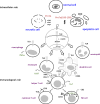
Fig. 1
Fig. 1
A proposed scenario explaining how proTα exerts its dual (intra- and extracellular) biologic effect. In a normal cell, proTα is mainly localized in the cell nucleus, where it regulates gene expression and cell proliferation. Under abnormal conditions, cells responding to danger signals die via necrosis or apoptosis. During necrosis, cell components, such as intact proTα, are released extracellularly, due to cell membrane disruption. During apoptosis, proTα is relocalized in the cytoplasm, where its carboxy-terminus is cleaved by caspases. ProTα’s truncation generates the immunoactive peptide, proTα(100–109), which polymerizes into β-sheet structures and is excreted from the cell. Extracellularly, both proTα and proTα(100–109) activate innate-immunity cells, for example, macrophages, monocytes, DC and neutrophils via TLR-ligation and signaling. Stimulation of monocytes and DC enhances antigen presentation and strengthens their synapsis with T cells. DC stimulate T cell proliferation and cytokine production, thus providing a favorable environment for enhancing specific and non-specific cytotoxicity. Consequently, effector cells produce lytic molecules (e.g., perforin) and upregulate adhesion-molecule expression (e.g., CD2). In the presence of specific antigens, CD8+ T cell cytotoxicity is enhanced. formula image secretion of cytokine; formula image stimulation of proliferation

Similar articles

Cited by

References

    1. Miller JF. Immunological function of the thymus. Lancet. 1961;2:748–749. doi: 10.1016/S0140-6736(61)90693-6. - DOI - PubMed
    1. Good RA, Dalmasso AP, Martinez C, Archer OK, Pierce JC, Papermaster BW. The role of the thymus in development of immunologic capacity in rabbits and mice. J Exp Med. 1962;116:773–796. doi: 10.1084/jem.116.5.773. - DOI - PMC - PubMed
    1. Aisenberg AC, Wilkes B. Partial immunological restoration of neonatally thymectomized rats with thymus-containing diffusion chambers. Nature. 1965;205:716–717. doi: 10.1038/205716a0. - DOI - PubMed
    1. Berthiaume F, Aparicio CL, Eungdamrong J, Yarmush ML. Age- and disease-related decline in immune function: an opportunity for “thymus-boosting” therapies. Tissue Eng. 1999;5:499–514. doi: 10.1089/ten.1999.5.499. - DOI - PubMed
    1. Schulof RS. Thymic peptide hormones: basic properties and clinical applications in cancer. Crit Rev Oncol Hematol. 1985;3:309–376. doi: 10.1016/S1040-8428(85)80035-4. - DOI - PubMed

Publication types

LinkOut - more resources