. 2012 Feb 26;11(4):316-22.
doi: 10.1038/nmat3253.

Self-assembled RNA interference microsponges for efficient siRNA delivery

Affiliations

Self-assembled RNA interference microsponges for efficient siRNA delivery

Jong Bum Lee et al. Nat Mater. .

Abstract

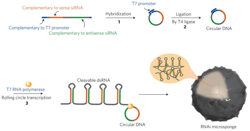
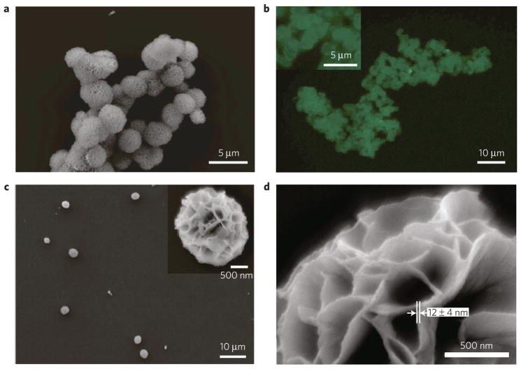
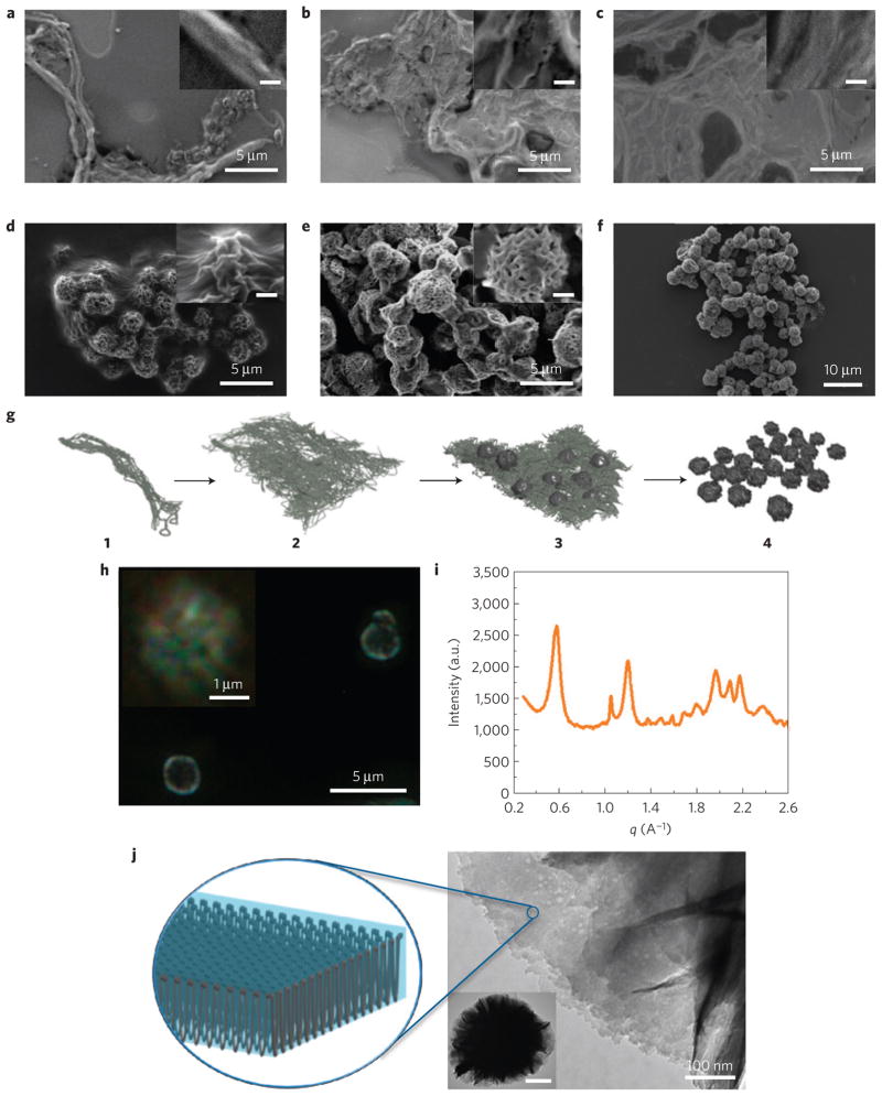
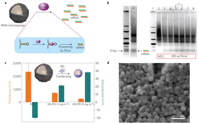
The encapsulation and delivery of short interfering RNA (siRNA) has been realized using lipid nanoparticles, cationic complexes, inorganic nanoparticles, RNA nanoparticles and dendrimers. Still, the instability of RNA and the relatively ineffectual encapsulation process of siRNA remain critical issues towards the clinical translation of RNA as a therapeutic. Here we report the synthesis of a delivery vehicle that combines carrier and cargo: RNA interference (RNAi) polymers that self-assemble into nanoscale pleated sheets of hairpin RNA, which in turn form sponge-like microspheres. The RNAi-microsponges consist entirely of cleavable RNA strands, and are processed by the cell's RNA machinery to convert the stable hairpin RNA to siRNA only after cellular uptake, thus inherently providing protection for siRNA during delivery and transport to the cytoplasm. More than half a million copies of siRNA can be delivered to a cell with the uptake of a single RNAi-microsponge. The approach could lead to novel therapeutic routes for siRNA delivery.


Conflict of interest statement

The authors declare no competing financial interests.

Figures

Figure 1
Figure 1. Schematic showing the process of rolling circle transcription (RCT) for the self-assembled RNAi-microsponge
To perform RCT (ref. 35), circular DNA needs to be synthesized first (see Supplementary Information). Linear single-stranded DNA that includes antisense and sense sequences of anti-luciferase siRNA is hybridized with an equal molar amount of short DNA strands containing the T7 promoter sequence. The nick in the circular DNA was chemically closed by T4 DNA ligase. By RCT of the closed circular DNA, multiple tandem repeats of hairpin RNA structures from both antisense and sense sequences are generated to be able to form spherical sponge-like structures.
Figure 2
Figure 2. Characterization of the RNAi-microsponge
a, SEM image of RNAi-microsponges. b, Fluorescence microscope image of RNAi-microsponges after staining with SYBR II, an RNA-specific dye. c, d, SEM images of RNAi-microsponges after sonication. Low-magnification image of an RNAi-microsponges (c). High-magnification image of an RNAi-microsponge (d).
Figure 3
Figure 3. Formation of sponge-like spherical structures purely with RNA strands
ae, SEM images of RNA products of time-dependent RCT at 37 °C for 1 h (a), 4 h (b), 8 h (c), 12 h (d) and 16 h (e). Inset: scale bar, 500 nm. f, Image of mature RNAi-microsponges after 20 h RCT. g, Schematic of the formation of RNAi-microsponges. The spherical sponge-like structure is formed through a series of preliminary structures. A tandem copy of RNA strands from the RCT reaction are entangled and twisted into a fibre-like structure 1. As the RNA strands grow, they begin to organize into lamellar sheets that gradually become thicker 2; as the internal structure of the sheets begins to get very dense, some of the RNA sheets begin to grow in the z direction, possibly owing to the limited packing area for the RNA polymer as it is produced by the reaction. This process could generate a wrinkled semi-spherical structure on the sheet 3. Finally, the entire structure begins to pinch off to form individual particles consisting of gathered RNA sheets 4. h, Polarized optical microscopy of RNAi-microsponges. i, X-ray diffraction pattern of RNAi-microsponges. j, TEM images of RNAi-microsponges and schematic representation of the proposed crystal-like ordered structure of an RNA sheet in the microsponge. Inset: scale bar, 500 nm.
Figure 4
Figure 4. Generating siRNA from RNAi-microsponges by the RNAi pathway, and condensing RNAi-microsponges for transfection
a, Schematic of the generation of siRNA from RNAi-microsponges by Dicer in the RNAi pathway. b, Gel-electrophoresis result after Dicer reaction. On the left, lanes 1 and 2 indicate double-stranded RNA ladder and RNAi-microsponges (MS) after treatment with Dicer (1 unit) for 36 h, respectively. On the right, lanes 1 and 2 indicate double-stranded RNA ladder and RNAi-microsponges without Dicer treatment. Lanes 3–8 correspond to 12 h, 24 h, 36 h and 48 h reaction with 1 unit of Dicer, and 36 h reaction with 1.25 and 1.5 units of Dicer, respectively. Increasing the amount of Dicer did not help to generate more siRNA (lane 7 and 8 of b). The amount of generated siRNA from RNAi-microsponges was quantified relative to double-stranded RNA standards. 21% of the cleavable double-stranded RNA was actually diced to siRNA because Dicer also produced the two or three repeat RNA units. The results suggest the possibility that in a more close-packed self-assembled structure, some portion of the RNA is not as readily accessed by Dicer. c, Particle size and zeta potential before and after condensing RNAi-microsponges with PEI. d, SEM image of further condensed RNAi-microsponges with PEI. The size of the RNAi-microsponges was significantly reduced by linear PEI because the RNAi-microsponges with high charge density would be more readily complexed with oppositely charged polycations. The porous structure of the RNAi-microsponges disappeared following condensation.
Figure 5
Figure 5. Transfection and gene silencing effect
a, Intracellular uptake of red fluorescent dye-labelled RNAi-microsponges without PEI (top) and RNAi-microsponge/PEI (bottom). To confirm the cellular transfection of RNA particles, both types of particles, labelled for red fluorescence, were incubated with T22 cells. Fluorescence labelled RNAi-microsponges without a PEI outer layer showed relatively less cellular uptake by the cancer cell line (T22 cells) suggesting that the larger size and strong net negative surface charge of RNAi-microsponges probably prevents cellular internalization. Inset: scale bar, 5 μm. b, Suppression of luciferase expression by siRNA, Lipofectamine complexed with siRNA (siRNA/Lipo), siRNA complex with PEI (siRNA/PEI), RNAi-microsponge (RNAi-MS), and RNAi-microsponge condensed by PEI (RNAi-MS/PEI). The same amount of siRNA is theoretically produced from RNAi-microsponges at the concentration in parentheses. c, In vivo knockdown of firefly luciferase by RNAi-MS/PEI. Optical images of tumours after intratumoral injection of RNAi-MS/PEI into the left tumour of a mouse and PEI solution only as a control into the right tumour of the same mouse.

Comment in

  • siRNA delivery: Loaded-up microsponges.
    Grabow WW, Jaeger L. Grabow WW, et al. Nat Mater. 2012 Mar 22;11(4):268-9. doi: 10.1038/nmat3286. Nat Mater. 2012. PMID: 22437781 No abstract available.
  • siRNA as a sponge.
    Baker M. Baker M. Nat Methods. 2012 Apr;9(4):327. doi: 10.1038/nmeth.1965. Nat Methods. 2012. PMID: 22563602 No abstract available.

Similar articles

Cited by

References

    1. Semple SC, et al. Rational design of cationic lipids for siRNA delivery. Nature Biotechnol. 2010;28:172–176. - PubMed
    1. Peer D, Park EJ, Morishita Y, Carman CV, Shimaoka M. Systemic leukocyte-directed siRNA delivery revealing cyclin D1 as an anti-inflammatory target. Science. 2008;319:627–630. - PMC - PubMed
    1. Mok H, Lee SH, Park JW, Park TG. Multimeric small interfering ribonucleic acid for highly efficient sequence-specific gene silencing. Nature Mater. 2010;9:272–278. - PubMed
    1. Liu L, et al. Self-assembled cationic peptide nanoparticles as an efficient antimicrobial agent. Nature Nanotechnol. 2009;4:457–463. - PubMed
    1. Lee JS, et al. Gold, poly(β-amino ester) nanoparticles for small interfering RNA delivery. Nano Lett. 2009;9:2402–2406. - PMC - PubMed

Publication types

Substances