. 2012 May;191(1):65-78.
doi: 10.1534/genetics.112.139105. Epub 2012 Feb 23.

Homologous recombination via synthesis-dependent strand annealing in yeast requires the Irc20 and Srs2 DNA helicases

Affiliations

Homologous recombination via synthesis-dependent strand annealing in yeast requires the Irc20 and Srs2 DNA helicases

Tohru Miura et al. Genetics. 2012 May.

Abstract

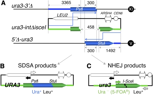
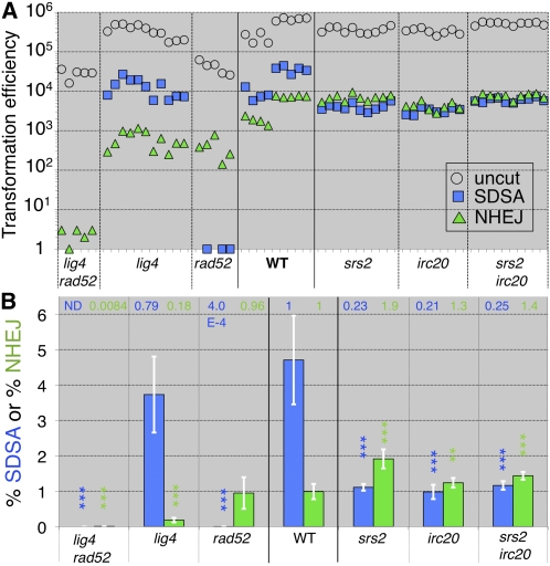
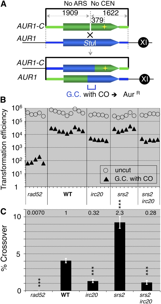
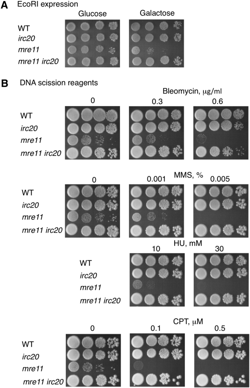
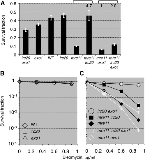
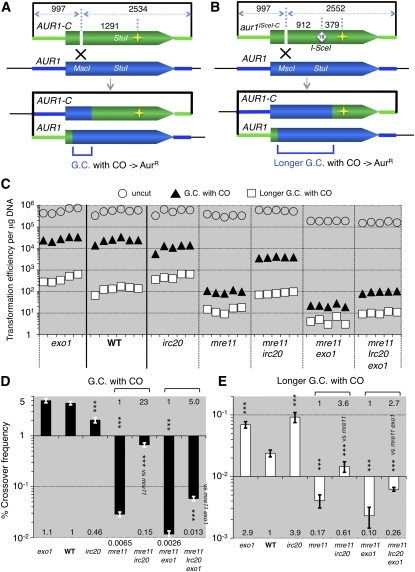
Synthesis-dependent strand-annealing (SDSA)-mediated homologous recombination replaces the sequence around a DNA double-strand break (DSB) with a copy of a homologous DNA template, while maintaining the original configuration of the flanking regions. In somatic cells at the 4n stage, Holliday-junction-mediated homologous recombination and nonhomologous end joining (NHEJ) cause crossovers (CO) between homologous chromosomes and deletions, respectively, resulting in loss of heterozygosity (LOH) upon cell division. However, the SDSA pathway prevents DSB-induced LOH. We developed a novel yeast DSB-repair assay with two discontinuous templates, set on different chromosomes, to determine the genetic requirements for somatic SDSA and precise end joining. At first we used our in vivo assay to verify that the Srs2 helicase promotes SDSA and prevents imprecise end joining. Genetic analyses indicated that a new DNA/RNA helicase gene, IRC20, is in the SDSA pathway involving SRS2. An irc20 knockout inhibited both SDSA and CO and suppressed the srs2 knockout-induced crossover enhancement, the mre11 knockout-induced inhibition of SDSA, CO, and NHEJ, and the mre11-induced hypersensitivities to DNA scissions. We propose that Irc20 and Mre11 functionally interact in the early steps of DSB repair and that Srs2 acts on the D-loops to lead to SDSA and to prevent crossoverv.


Figures

Figure 1
Figure 1
DSB repair pathways. (A) Holliday-junction (HJ)-mediated homologous recombination. This pathway involves the resolution of two Holliday junctions. A1: Both ends at the DSB are unwound and exonucleolytically degraded, resulting in the formation of a 3′-single-strand (ss)-tailed DNA. A2: One 3′-ss-tailed DNA invades into the homologous DNA, and a D-loop with a short heteroduplex is formed. A3: The heteroduplex region is expanded in the 5′ direction by the strand-exchange reaction, and a Holliday junction is formed. A4: From the 3′ end of the heteroduplex, DNA-repair synthesis is primed. Strand exchange occurs at the other end of the DSB, and the second Holliday junction is formed. A5 or A6: One of two types of products, gene conversion or gene conversion associated with crossover, is generated depending on how the two Holliday junctions are resolved. The ends-in gene targeting produces the targeted integrant (A6) through the association of gene conversion with crossover, but does not through gene conversion without crossover (A5). (B) Synthesis-dependent strand-annealing (SDSA)-mediated homologous recombination. This pathway is associated with D-loop migration. B1: This intermediate is identical to A1. B2: This intermediate is identical to A2. B3: Immediately after D-loop formation, repair synthesis is primed from the 3′ end of the heteroduplex DNA. While DNA synthesis continues, the rear heteroduplex region is unwound. B4: As DNA synthesis proceeds to the homologous region with the other DSB end, the newly synthesized strand is detached. B5: The detached strand is reannealed to the other ss-tailed region, and the gap is filled. The arrow from A4 to B5 indicates that two heteroduplex molecules linked by a double-Holliday junction (A4) are resolved into separate duplexes (B5), by migrating the two Holliday junctions into the center. (C) Nonhomologous end-joining (NHEJ) pathway. C1: Both ends of a DSB are unwound. C2: Two unwound ends interact with each other through microhomology. C3: Immediately after this interaction, the two ends are ligated. C4: The end-joining product is formed.
Figure 2
Figure 2
SDSA/NHEJ assay system. (A) The different ura3 template alleles and the I-SceI-cut plasmid bearing the ura3 recipient allele with a gap. One ura3 template allele is 5′Δ-ura3, in which the region from the promoter to the 39th codon was deleted in the URA3 locus on the 5th chromosome. The other ura3 template allele is ura3-3′Δ, which is the ectopic allele containing the region from the URA3 promoter to the 139th codon, located within the AUR1 locus on the 11th chromosome. These template alleles share 300 bp of internal homology. The ura3 recipient allele is ura3-intΔisceI, which lacks the 458-bp region from the PstI site to the StuI site. As this gap is sealed with the 18-bp I-SceI sequence, I-SceI cleavage produces the double-strand gap. The plasmid bearing ura3-intΔisceI has ARSH4, CEN6, and LEU2 selection marker gene. (B) SDSA products are obtained as Ura+ Leu+ transformants bearing the plasmid with the URA3 allele, which is repaired via SDSA using the two templates. The reversion of the 458-bp gap was verified by PCR for the entire region of the plasmid DNA with a pair of primers, PRI271 (solid arrow) and PRI274 (open arrow), and PstI/StuI digestion (Figure S1A). (C) NHEJ products are obtained as Ura- (5-FOAR) Leu+ transformants bearing the plasmid with an ura3 allele. The retention or deletion of the I-SceI sequence was detected by PCR for the plasmid DNA with the primers PRI271 (solid arrow) and PRI274 (open arrow) or with another pair of primers, PRI231 (green arrow) and PRI277 (gray arrow), I-SceI digestion (Figure S1B), and sequence determination (Table 2).
Figure 3
Figure 3
Frequencies of SDSA and NHEJ events in lig4, rad52, srs2, and irc20-deficient mutants. (A) Leu+ transformation efficiencies per microgram with the uncut plasmid are plotted as the transformation competencies of competent cell suspensions (open symbols). Ura+ Leu+ and 5-FOAR Leu+ transformation efficiencies per microgram with the I-SceI cut plasmid are plotted as SDSA progeny (blue symbols) and NHEJ progeny (green symbols), respectively. (B) The normalized SDSA (blue bar) and NHEJ (green bar) frequencies (%) were calculated from the transformation efficiencies (Materials and Methods), and were plotted with the standard deviations (SD) (white lines). The SDSA and the NHEJ frequencies (%) (mean ±SD) and the numbers of transformation of the indicated strains are shown in Table S1. ND indicates that SDSA progeny was not detected. The results from the t-test (vs. wild-type) are shown as follows: **, P < 0.01; ***, P < 0.001 (blue for SDSA; green for NHEJ).
Figure 4
Figure 4
Frequency of crossover events in srs2 and irc20-deficient mutants. (A) The AUR1-C plasmid DNA is cleaved by the StuI enzyme, whose site is 379 bp away from the AUR1-C dominant mutation. The resultant 5′ end and 3′ end share 1909-bp homology and 1622-bp homology with the AUR1 gene (chromosome XI), respectively. (B) AurR transformation efficiencies per microgram with the uncut pRS315-AurR plasmid are plotted as the transformation competencies of competent cell suspensions (open symbols). AurR transformation efficiencies per microgram with the StuI cut plasmid are plotted as the numbers of crossover progeny (solid symbols). (C) The normalized frequencies of crossover events associated with gene conversion (black bar) were calculated from the transformation efficiencies (Materials and Methods), and the means were plotted with the SD (white line). The means (±SD) (%) of the wild-type strain (WT) and the indicated knockout strains are as follows: WT 4.1 (±0.29); rad52 0.028 (±0.012); srs2 9.3 (±0.80); irc20 1.3 (±0.21); srs2 irc20 1.1 (±0.25). The results from the t-test (vs. wild type) are shown as follows: ***, P < 0.001.
Figure 5
Figure 5
Frequencies of SDSA and NHEJ events in mre11- and irc20-deficient mutants. (A) Leu+ transformation efficiencies per microgram with the uncut plasmid are plotted as the transformation competencies of competent cell suspensions (open symbols). Ura+ Leu+ and 5FOAR Leu+ transformation efficiencies per microgram with the I-SceI cut plasmid are plotted as SDSA progeny (blue symbols) and NHEJ progeny (green symbols), respectively. (B) The normalized SDSA (blue bar) and NHEJ (green bar) frequencies (%) were calculated from the transformation efficiencies (Materials and Methods), and the means were plotted with the SD (white lines). The SDSA and the NHEJ frequencies (%) (mean ±SD) and the numbers of transformation of the indicated strains are shown in Table S1. The results from the t-test (vs. wild type except for vs. mre11) are shown as follows: *, P < 0.05; ***, P < 0.001 (blue for SDSA; green for NHEJ).
Figure 6
Figure 6
Effects of mre11 and irc20 mutations on the repair of DNA scission-induced DSBs. Each of the spots is a serial 10-fold dilution from a culture of the strain with the indicated genotype. (A) EcoRI expression was induced on minimal plates containing galactose. (B) The DNA scission reagents bleomycin, MMS, HU, and CPT were added at the indicated concentrations to YPD plates.
Figure 7
Figure 7
Effects of the exo1 mutation on the irc20 knockout-induced suppression of mre11 defects in DSB repair. (A) Repair of EcoRI-induced DSBs. The surviving fraction (Materials and Methods) was plotted along with the SD (vertical line). The number (n) of experiments for each strain is as follows: WT (6), irc20 (4), mre11 (9), mre11 irc20 (4), mre11 exo1 (4), mre11 irc20 exo1 (5), exo1 (4), and irc20 exo1 (4). (B\x{2013}C) Repair of bleomycin-induced DSBs. The surviving fraction (Materials and Methods) was plotted along with the SD (vertical line). The number (n) of experiments for each strain is as follows: WT (4), irc20 (4), mre11 (10), mre11 irc20 (4), mre11 exo1 (3), mre11 irc20 exo1 (6), exo1 (6), and irc20 exo1 (6).
Figure 8
Figure 8
Frequency of the crossover events associated with longer-tract gene conversion in mre11 and irc20-deficient mutants. (A) The pAUR101 plasmid DNA was digested by MscI. The AUR1-C dominant mutation is 1291 bp away from the DSB end. The resultant 5′ and 3′ ends share 997-bp and 2534-bp homologies with the AUR1 gene on the 11th chromosome, respectively. The targeted integration by a crossover event produces a tandem duplication of AUR1-C and AUR1 to confer AurR. (B) The pAUR101-I-SceI plasmid DNA, possessing the 18-bp (I-SceI sequence) insertion at the StuI site, digested by MscI. This insertion is 912 bp away from the DSB end, and the AUR1-C dominant mutation is 379 bp away from the insertion. The resultant 5′ and 3′ ends share 997-bp and 2552-bp homologies with the AUR1 gene on the 11th chromosome, respectively. The targeted integration by a crossover event with associated longer gene conversion produces a tandem duplication of AUR1-C and AUR1 to confer AurR. (C) AurR transformation efficiencies per microgram of the uncut pRS315-AurR plasmid are plotted as the transformation competencies of competent cell suspensions (open circles). AurR transformation efficiencies per microgram of the MscI-cut pAUR101 plasmid are plotted as the numbers of crossover progeny (solid triangles). AurR transformation efficiencies per microgram of the MscI-cut pAUR101-I-SceI plasmid are plotted as the numbers of crossover progeny (open squares). (D) The normalized crossover frequencies (black bar) were calculated from the transformation efficiencies (Materials and Methods), and the means were plotted along with the SD (white line). The means (±SD) (%) of the wild-type strain (WT) and the indicated knockout strains are as follows: WT 4.4 (±0.32); irc20 2.0 (±0.25); mre11 0.029 (±0.0030); mre11 irc20 0.66 (±0.063); mre11 exo1 0.012 (±0.0018); mre11 irc20 exo1 0.058 (±0.0058); exo1 4.9 (±0.47). (E) The normalized frequencies of crossover associated with longer-tract gene conversion (white bar) were calculated from the transformation efficiencies (Materials and Methods), and the means were plotted along with the SD (black line). The means (±SD) (%) of the wild-type strain (WT) and the indicated knockout strains are as follows: WT 0.024 (±0.0030); irc20 0.092 (±0.017); mre11 0.0041 (±0.00098); mre11 irc20 0.015 (±0.0028); mre11 exo1 0.0023 (±0.00086); mre11 irc20 exo1 0.0062 (±0.00045); exo1 0.069 (±0.0084). The results from the t-test (vs. wild type except for vs. mre11) are shown as follows: *, P < 0.05; ***, P < 0.001.

Similar articles

Cited by

References

    1. Adams M. D., McVey M., Sekelsky J. J., 2003. Drosophila BLM in double-strand break repair by synthesis-dependent strand annealing. Science 299: 265–267. - PubMed
    1. Aguilera A., Klein H. L., 1988. Genetic control of intrachromosomal recombination in Saccharomyces cerevisiae. I. Isolation and genetic characterization of hyper-recombination mutations. Genetics 119: 779–790. - PMC - PubMed
    1. Alvaro D., Lisby M., Rothstein R., 2007. Genome-wide analysis of Rad52 foci reveals diverse mechanisms impacting recombination. PLoS Genet. 3: e228. - PMC - PubMed
    1. Bignon Y.-J., 2004. Biological basis of cancer predisposition, pp. 11–20 in Genetic Predisposition to Cancer, edited by Eeles R. A., Easton D. F., Ponder B. A. J., Eng C. Arnold, New York.
    1. Boulton S. J., Jackson S. P., 1996a. Identification of a Saccharomyces cerevisiae Ku80 homologue: roles in DNA double strand break rejoining and in telomeric maintenance. Nucleic Acids Res. 24: 4639–4648. - PMC - PubMed

Publication types

MeSH terms

Substances