

Fusion of a novel genetically engineered chitosan affinity protein and green fluorescent protein for specific detection of chitosan in vitro and in situ
- PMID: 22367086
- PMCID: PMC3346462
- DOI: 10.1128/AEM.07506-11
Fusion of a novel genetically engineered chitosan affinity protein and green fluorescent protein for specific detection of chitosan in vitro and in situ
Abstract
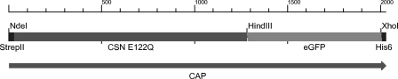
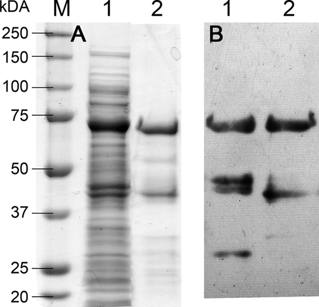
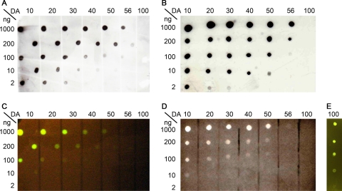
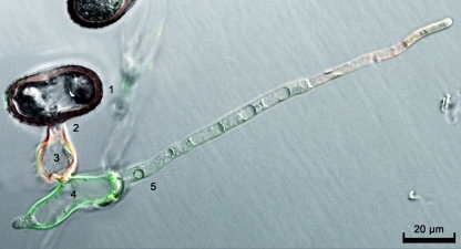
Chitin is the second most abundant polysaccharide, present, e.g., in insect and arthropod exoskeletons and fungal cell walls. In some species or under specific conditions, chitin appears to be enzymatically de-N-acetylated to chitosan-e.g., when pathogenic fungi invade their host tissues. Here, the deacetylation of chitin is assumed to represent a pathogenicity mechanism protecting the fungus from the host's chitin-driven immune response. While highly specific chitin binding lectins are well known and easily available, this is not the case for chitosan-specific probes. This is partly due to the poor antigenicity of chitosan so that producing high-affinity, specific antibodies is difficult. Also, lectins with specificity to chitosan have been described but are not commercially available, and our attempts to reproduce the findings were not successful. We have, therefore, generated a fusion protein between a chitosanase inactivated by site-directed mutagenesis, the green fluorescent protein (GFP), and StrepII, as well as His(6) tags for purification and detection. The recombinant chitosan affinity protein (CAP) expressed in Escherichia coli was shown to specifically bind to chitosan, but not to chitin, and the affinity increased with decreasing degree of acetylation. In vitro, CAP detection was possible either based on GFP fluorescence or using Strep-Tactin conjugates or anti-His(5) antibodies. CAP fluorescence microscopy revealed binding to the chitosan exposing endophytic infection structures of the wheat stem rust fungus, but not the chitin exposing ectophytic infection structures, verifying its suitability for in situ chitosan staining.
Figures
Similar articles
-
Characterization of antifungal activity of the GH-46 subclass III chitosanase from Bacillus circulans MH-K1.Antonie Van Leeuwenhoek. 2013 Nov;104(5):737-48. doi: 10.1007/s10482-013-9982-5. Epub 2013 Jul 27. Antonie Van Leeuwenhoek. 2013. PMID: 23892828
-
Amino Groups of Chitosan Are Crucial for Binding to a Family 32 Carbohydrate Binding Module of a Chitosanase from Paenibacillus elgii.J Biol Chem. 2016 Sep 2;291(36):18977-90. doi: 10.1074/jbc.M116.721332. Epub 2016 Jul 12. J Biol Chem. 2016. PMID: 27405759 Free PMC article.
-
A Recombinant Fungal Chitin Deacetylase Produces Fully Defined Chitosan Oligomers with Novel Patterns of Acetylation.Appl Environ Microbiol. 2016 Oct 27;82(22):6645-6655. doi: 10.1128/AEM.01961-16. Print 2016 Nov 15. Appl Environ Microbiol. 2016. PMID: 27590819 Free PMC article.
-
Various methods for determination of the degree of N-acetylation of chitin and chitosan: a review.J Agric Food Chem. 2009 Mar 11;57(5):1667-76. doi: 10.1021/jf803001m. J Agric Food Chem. 2009. PMID: 19187020 Review.
-
Chitin--the undisputed biomolecule of great potential.Crit Rev Food Sci Nutr. 2003;43(1):61-87. doi: 10.1080/10408690390826455. Crit Rev Food Sci Nutr. 2003. PMID: 12587986 Review.
Cited by
-
In-vitro Detection of Phytopathogenic Fungal Cell Wall by Polyclonal Sera Raised Against Trimethyl Chitosan Nanoparticles.Int J Nanomedicine. 2019 Dec 20;14:10023-10033. doi: 10.2147/IJN.S220488. eCollection 2019. Int J Nanomedicine. 2019. PMID: 31908457 Free PMC article.
-
The principles and applications of avidin-based nanoparticles in drug delivery and diagnosis.J Control Release. 2017 Jan 10;245:27-40. doi: 10.1016/j.jconrel.2016.11.016. Epub 2016 Nov 16. J Control Release. 2017. PMID: 27865853 Free PMC article. Review.
-
Complete Genome Sequence of Sporisorium scitamineum and Biotrophic Interaction Transcriptome with Sugarcane.PLoS One. 2015 Jun 12;10(6):e0129318. doi: 10.1371/journal.pone.0129318. eCollection 2015. PLoS One. 2015. PMID: 26065709 Free PMC article.
-
Purification and characterization of exo-β-1,4-glucosaminidase produced by chitosan-degrading fungus, Penicillium sp. IB-37-2A.World J Microbiol Biotechnol. 2019 Jan 17;35(2):18. doi: 10.1007/s11274-019-2590-4. World J Microbiol Biotechnol. 2019. PMID: 30656484
-
Chitin and chitosan remodeling defines vegetative development and Trichoderma biocontrol.PLoS Pathog. 2020 Feb 20;16(2):e1008320. doi: 10.1371/journal.ppat.1008320. eCollection 2020 Feb. PLoS Pathog. 2020. PMID: 32078661 Free PMC article.
References
-
- Adachi W, et al. 2004. Crystal structure of family GH-8 chitosanase with subclass II specificity from Bacillus sp. K17. J. Mol. Biol. 343:785–795 - PubMed
-
- Alzari PM, Souchon H, Dominguez R. 1996. The crystal structure of endoglucanase CelA, a family 8 glycosyl hydrolase from Clostridium thermocellum. Structure 4:265–275 - PubMed
-
- Baldo BA, Barnett D, Lee JW. 1984. Lectins as cytochemical probes of the developing wheat-grain.V. Demonstration of separate polysaccharides containing N-acetyl-d-glucosamine and d-galactose in nucellar epidermal-cell walls. Aust. J. Plant Physiol. 11:179–190
-
- Benhamou N, Asselin A. 1989. Attempted localization of a substrate for chitinases in plant-cells reveals abundant N-acetyl-d-glucosamine residues in secondary walls. Biol. Cell 67:341–350
MeSH terms
Substances
LinkOut - more resources
Full Text Sources
Other Literature Sources
Miscellaneous
