

Ultrastructural changes in the cemento-enamel junction after vital tooth bleaching with fluoride and fluoride-free agents - a pilot study
- PMID: 22367139
- PMCID: PMC3560748
- DOI: 10.12659/msm.882502
Ultrastructural changes in the cemento-enamel junction after vital tooth bleaching with fluoride and fluoride-free agents - a pilot study
Abstract
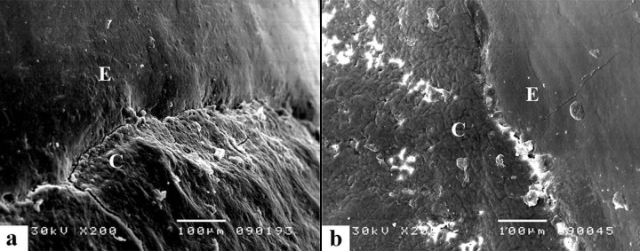
Background: The impact of bleaching on the cemento-enamel junction (CEJ) is not well known. Due to frequent sensitivity of the cervical region of teeth after the vital bleaching, the aim of the present study was to evaluate the morphological features of the CEJ of human teeth after application of fluoridated and fluoride-free bleaching agents, as well as post-bleaching fluoridation treatment, by scanning electron microscopy (SEM) analysis.
Material/methods: Thirty-five extracted permanent human teeth were longitudinally cut, yielding 70 specimens. Thirty specimens were randomly divided into the 3 experimental groups, and 20 specimens, were used as (2) control groups, each: negative (untreated) control group; positive control group treated with 35% hydrogen peroxide; experimental group 1, bleaching with 10% carbamide peroxide (CP); experimental group 2, treatment with a mixture of 10% CP and fluoride; and experimental group 3, treatment with 10% CP and 2% sodium fluoride gel applied 30 minutes after bleaching. Experimental groups were treated 8 h per day for 14 days. The samples were examined by SEM.
Results: The bleaching materials tested caused morphological changes to the surface of the CEJ. There was a statistically significant difference between experimental groups (Kruskal Wallis Test chi-square=11,668; p<0.005). Mean value of experimental group 2 scores showed statistically significant difference from groups 1 and 3.
Conclusions: Bleaching gel with fluorides does not significantly change morphological appearance of the CEJ and represents a better choice than the hard tissue fluoridation process after bleaching.
Figures


Similar articles
-
The application of casein phosphopeptide and amorphous calcium phosphate with fluoride (CPP-ACPF) for restoring mineral loss after dental bleaching with hydrogen or carbamide peroxide: An in vitro study.Ann Anat. 2019 Sep;225:48-53. doi: 10.1016/j.aanat.2019.05.005. Epub 2019 Jul 2. Ann Anat. 2019. PMID: 31271888
-
Effect of bleaching on the cemento-enamel junction.Am J Dent. 2007 Aug;20(4):245-9. Am J Dent. 2007. PMID: 17907488 Clinical Trial.
-
Clinical comparative study of the effectiveness of and tooth sensitivity to 10% and 20% carbamide peroxide home-use and 35% and 38% hydrogen peroxide in-office bleaching materials containing desensitizing agents.Oper Dent. 2012 Sep-Oct;37(5):464-73. doi: 10.2341/11-337-C. Epub 2012 May 18. Oper Dent. 2012. PMID: 22616927 Clinical Trial.
-
Effect of fluoride containing bleaching agents on enamel surface properties.J Dent. 2008 Sep;36(9):718-25. doi: 10.1016/j.jdent.2008.05.003. Epub 2008 Jun 24. J Dent. 2008. PMID: 18573586
-
Effect of different peroxide bleaching regimens and subsequent fluoridation on the hardness of human enamel and dentin.J Prosthet Dent. 2004 Oct;92(4):337-42. doi: 10.1016/j.prosdent.2004.07.019. J Prosthet Dent. 2004. PMID: 15507905
References
-
- Arambawata K, Peiris R, Nanayakkara Morphology of the cemento-enamel junction in premolar teeth. J Oral Sci. 2009;51:623–27. - PubMed
-
- Zantner C, Beheim-Schwarzbach N, Neumann K, Kielbassa AM. Surface microhardness of enamel after different home bleaching procedures. Dent Mater. 2007;23:243–50. - PubMed
-
- Attin T, Schmidlin PR, Wegehaupt F, Wiegand A. Influence of study design on on the impact of bleaching agents on dental enamel microhardness: a review. Dent Mater. 2009;25:143–57. - PubMed
-
- Joiner A. Review of the effects of peroxide on enamel and dentin properties. J Dent. 2007;35:882–96. - PubMed
Publication types
MeSH terms
Substances
LinkOut - more resources
Full Text Sources
Miscellaneous
