. 2012 Apr 13;287(16):13194-205.
doi: 10.1074/jbc.M112.339655. Epub 2012 Feb 24.

Structure and mechanistic insights into novel iron-mediated moonlighting functions of human J-protein cochaperone, Dph4

Affiliations

Structure and mechanistic insights into novel iron-mediated moonlighting functions of human J-protein cochaperone, Dph4

Anushikha Thakur et al. J Biol Chem. .

Abstract

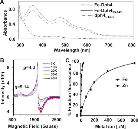
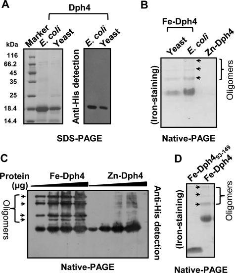
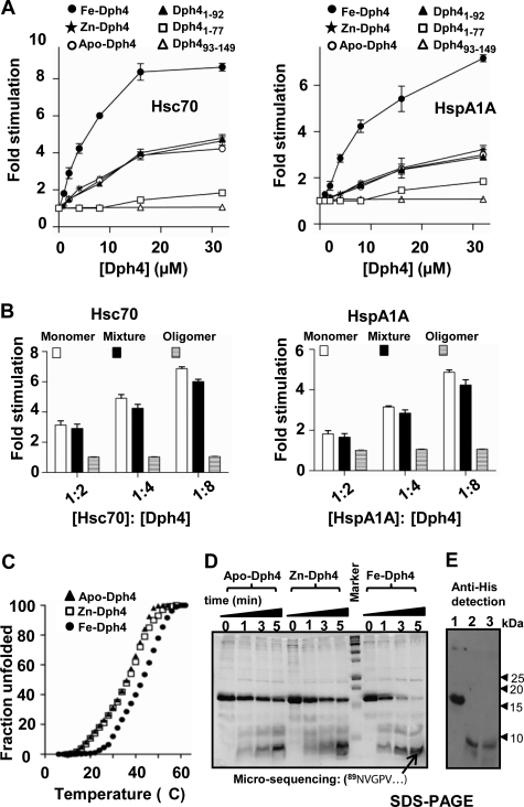
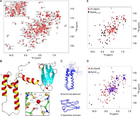
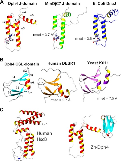
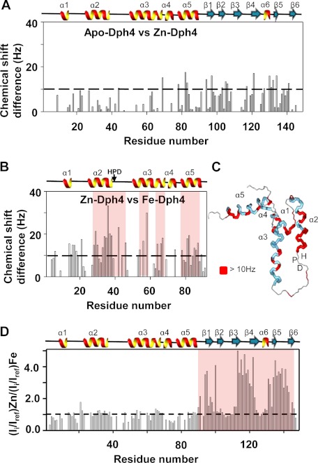
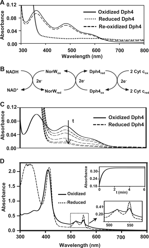
J-proteins are obligate cochaperones of Hsp70s and stimulate their ATPase activity via the J-domain. Although the functions of J-proteins have been well understood in the context of Hsp70s, their additional co-evolved "physiological functions" are still elusive. We report here the solution structure and mechanism of novel iron-mediated functional roles of human Dph4, a type III J-protein playing a vital role in diphthamide biosynthesis and normal development. The NMR structure of Dph4 reveals two domains: a conserved J-domain and a CSL-domain connected via a flexible linker-helix. The linker-helix modulates the conformational flexibility between the two domains, regulating thereby the protein function. Dph4 exhibits a unique ability to bind iron in tetrahedral coordination geometry through cysteines of its CSL-domain. The oxidized Fe-Dph4 shows characteristic UV-visible and electron paramagnetic resonance spectral properties similar to rubredoxins. Iron-bound Dph4 (Fe-Dph4) also undergoes oligomerization, thus potentially functioning as a transient "iron storage protein," thereby regulating the intracellular iron homeostasis. Remarkably, Fe-Dph4 exhibits vital redox and electron carrier activity, which is critical for important metabolic reactions, including diphthamide biosynthesis. Further, we observed that Fe-Dph4 is conformationally better poised to perform Hsp70-dependent functions, thus underlining the significance of iron binding in Dph4. Yeast Jjj3, a functional ortholog of human Dph4 also shows a similar iron-binding property, indicating the conserved nature of iron sequestration across species. Taken together, our findings provide invaluable evidence in favor of additional co-evolved specialized functions of J-proteins, previously not well appreciated.


Figures

FIGURE 1.
FIGURE 1.
Analysis of iron binding in Dph4. A, UV-visible absorption spectra of 200 μm Fe-Dph4, Fe-Dph4(93–149), and dph4C139S. B, EPR spectra of 0.6 mm Fe-Dph4 in solution recorded at different temperatures. The observed resonances are marked for their g values. C, titration of apo-Dph4 (2 μm) with Fe2+ as (NH4)2Fe(SO4)2·6H2O (in 200 μm ascorbic acid) and Zn2+ as ZnSO4·7H2O. The change in Trp fluorescence intensity (342 nm) of apo-Dph4 was monitored.
FIGURE 2.
FIGURE 2.
Oligomerization in Fe-Dph4. A, immunodetection of Dph4 purified from yeast. Purified protein was separated on SDS-PAGE and immunodetected by Western blotting using anti-His antibody. B, iron-specific staining of Dph4 purified from E. coli and yeast separated on native PAGE. C, Western blot of Fe-Dph4 and Zn-Dph4 separated on native-PAGE and immunodetected by anti-His antibody. Increasing amounts of protein (4–20 μg) were loaded as indicated. D, iron staining of Dph4(93–149) purified from E. coli separated on native PAGE. Oligomers are marked with arrows.
FIGURE 3.
FIGURE 3.
ATPase assays of Dph4. A, stimulation of Hsc70 (left) and HspA1A (right) by apo-Dph4, Zn-Dph4, Fe-Dph4, Dph4(1–76), Dph4(1–92), and Fe-Dph4(93–149). The preformed radiolabeled Hsc70-ATP (1 μm) and Hsp70A1A-ATP (1 μm) complexes were incubated with increasing concentrations of Fe-Dph4 or Zn-Dph4. ATP hydrolysis was monitored under single turnover conditions at different time intervals at 25 °C, and rates of hydrolysis were calculated for different concentrations. -Fold stimulation was calculated by setting the intrinsic ATP hydrolysis rate as 1. Error bars are derived from two independent sets of experiments. B, stimulation of Hsc70 (left) and HspA1A (right) by monomer and oligomers of Fe-Dph4. C, thermal denaturation curve of apo-, Zn-, and Fe-Dph4 using circular dichroism. The fractional change in ellipticity at 222 nm was monitored. D, trypsin digestion of Fe-, Zn-, and apo-Dph4. The digestion was analyzed at different time points as indicated. The marked band was subjected to N-terminal microsequencing. The sequence is indicated in the figure. E, anti-His immunoblot of trypsin-digested Fe-Dph4. Lane 1, Fe-Dph4; lane 2, Fe-Dph4 after 10 min of trypsin digestion; lane 3, Dph4(93–149).
FIGURE 4.
FIGURE 4.
Structural analysis of full-length Zn-Dph4. A, two-dimensional 15N-1H HSQC spectrum of Zn-Dph4 acquired at 1H resonance frequency of 700 MHz at 298 K. Sequence-specific resonance assignments are indicated by labels depicting the single-letter code for the amino acid followed by the residue number. Side chain amide signals of asparagines and glutamines are shown connected by horizontal lines. Peaks from residues undergoing conformational exchange are marked by asterisks. The overlapped region of the spectrum is expanded in the inset. B, ribbon diagram of representative structure of Zn-Dph4, Zn2+ (red) in tetrahedral coordination with Sγ (gold) (inset). The average Zn–Sγ distance is marked with dotted lines. C, superimposition of the 20 best structures of N-terminal J-domain (top) and C-terminal CSL-domain (bottom). D, overlay of two-dimensional 15N-1H HSQC spectrum of full-length Zn-Dph4 and Dph4(1–92). E, overlay of two-dimensional 15N-1H HSQC spectrum of Zn-Dph4 and Dph4(93–149).
FIGURE 5.
FIGURE 5.
Comparison of Dph4 subdomains with representative structures of similar folds. A, J-domain of Dph4, MmDjC7 (PDB code 1WJZ), and E. coli DnaJ (PDB code 1XBL) (top). B, CSL-domain of Dph4, human DESR1 (PDB code 2JR7) and yeast Kti11 (PDB code 1YOP) (middle). The r.m.s. deviations among the domains are indicated. The signature HPD motif of J-domains is represented in a ball and stick model. C, comparison of full-length human Dph4 and human HscB.
FIGURE 6.
FIGURE 6.
Chemical shift perturbation analysis of apo-, Zn-, and Fe-Dph4. Chemical shift differences between apo- and Zn-Dph4 (A) and Zn- and Fe-Dph4 (B) as a function of residues. Bars crossing 10 Hz (as shown by the dashed line) indicate significantly perturbed amino acid residues. C, chemical shift differences of >10 Hz (red) between Zn- and Fe-Dph4 mapped onto the protein structure. Helices and loops are indicated in blue and gray, respectively. D, plot showing the ratio of peak intensities in 15N-1H HSQC spectra of Zn-Dph4 and Fe-Dph4. Bars crossing the threshold (shown as a dotted line) indicate broadening due to paramagnetic effect of Fe3+. The absence of a bar indicates a missing assignment. The secondary structure of the protein is plotted at the top of each panel, and the HPD-motif is indicated. The perturbed regions are highlighted in red.
FIGURE 7.
FIGURE 7.
Redox and electron carrier properties of Fe-Dph4. A, UV-visible absorption spectra of 20 μm Fe-Dph4 reduced by 1.5 mm sodium dithionite in anaerobic conditions followed by reoxidation in air. B, scheme of in vitro electron carrier assay showing the transfer of electrons from NADH to cytochrome c via E. coli NorW and Fe-Dph4. C, anaerobic reduction of 18 μm Fe-Dph4 by 1 μm NorW at different time intervals after the addition of 400 μm NADH at 37 °C. D, demonstration of electron transfer from Fe-Dph4 (20 μm) to cytochrome c (300 μg/ml) in the presence of NorW (2 μm) and NADH (300 μm) under anaerobic conditions at 37 °C. Upper inset, Absorbance traces at 550 nm following the time course for reduction of 200 μg/ml cytochrome c by 5 μm Fe-Dph4 in the presence 1 μm NorW and 300 μm NADH. Lower inset, an expanded view of reduction of cytochrome c observed at 550 nm.
FIGURE 8.
FIGURE 8.
In vitro and in vivo functional analysis of Dph4. A, in vivo rescue of Δjjj3 phenotype by DPH4. All strains indicated contain DT plasmid under galactose regulation. Serial dilutions of wild type strain with empty vector (W303), Δjjj3 with empty vector (−), or 2μ-GPD vector expressing full-length JJJ3 (Jjj3) or full-length DPH4 (Dph4) were spotted on dropout media containing 2% glucose or galactose and incubated for 72 h at 30 °C. B, UV-visible absorption spectra of 200 μm Fe-Dph4 and iron-bound Jjj3 (Fe-Jjj3) purified from E. coli grown in iron-supplemented M9 medium recorded at 25 °C. Inset, nickel bead-bound Fe-Dph4 and iron-bound Jjj3 proteins. C, iron-bound Jjj3 separated on native-PAGE and stained for iron. D, immunodetection of iron-bound Jjj3 by anti-His antibody. Oligomers are marked with arrows.

Similar articles

Cited by

References

    1. Craig E. A., Huang P., Aron R., Andrew A. (2006) The diverse roles of J-proteins, the obligate Hsp70 co-chaperone. Rev. Physiol. Biochem. Pharmacol. 156, 1–21 - PubMed
    1. Bukau B., Weissman J., Horwich A. (2006) Molecular chaperones and protein quality control. Cell 125, 443–451 - PubMed
    1. Walsh P., Bursać D., Law Y. C., Cyr D., Lithgow T. (2004) The J-protein family. Modulating protein assembly, disassembly and translocation. EMBO Rep. 5, 567–571 - PMC - PubMed
    1. Mayer M. P., Brehmer D., Gässler C. S., Bukau B. (2001) Hsp70 chaperone machines. Adv. Protein Chem. 59, 1–44 - PubMed
    1. Mayer M. P., Bukau B. (2005) Hsp70 chaperones. Cellular functions and molecular mechanism. Cell Mol. Life Sci. 62, 670–684 - PMC - PubMed

Publication types

MeSH terms

Associated data