. 2012 Apr 13;287(16):12913-26.
doi: 10.1074/jbc.M111.331751. Epub 2012 Feb 24.

A novel cardiomyocyte-enriched microRNA, miR-378, targets insulin-like growth factor 1 receptor: implications in postnatal cardiac remodeling and cell survival

Affiliations

A novel cardiomyocyte-enriched microRNA, miR-378, targets insulin-like growth factor 1 receptor: implications in postnatal cardiac remodeling and cell survival

Ivana Knezevic et al. J Biol Chem. .

Abstract

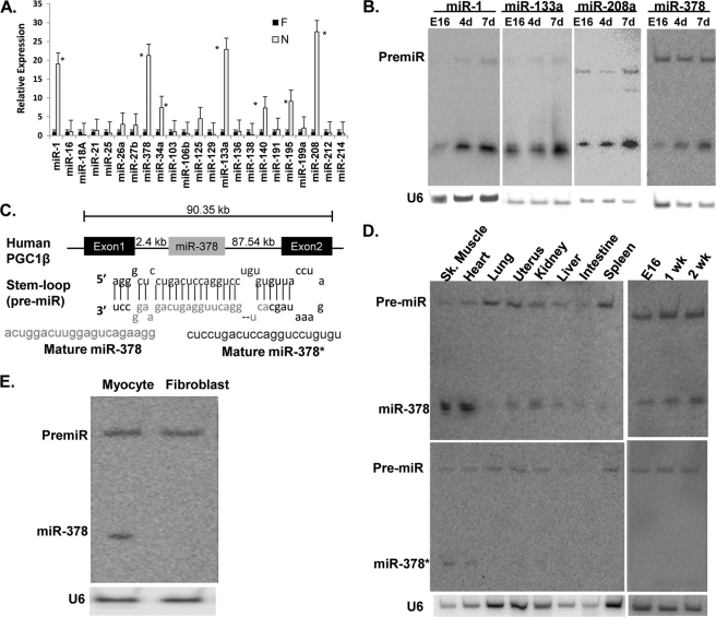
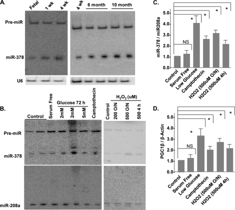
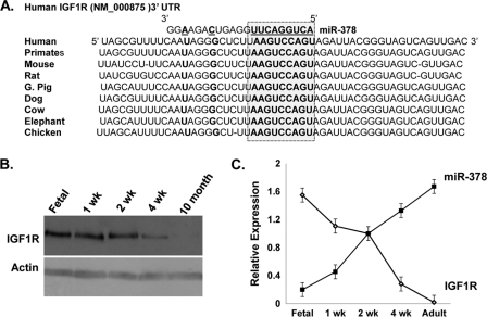
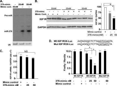
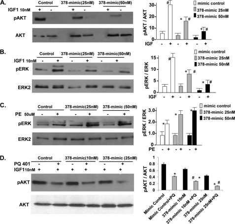
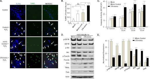
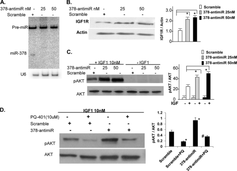
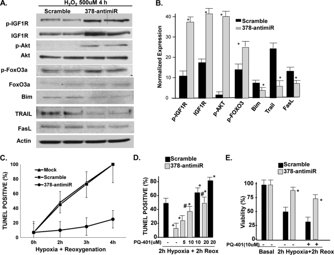
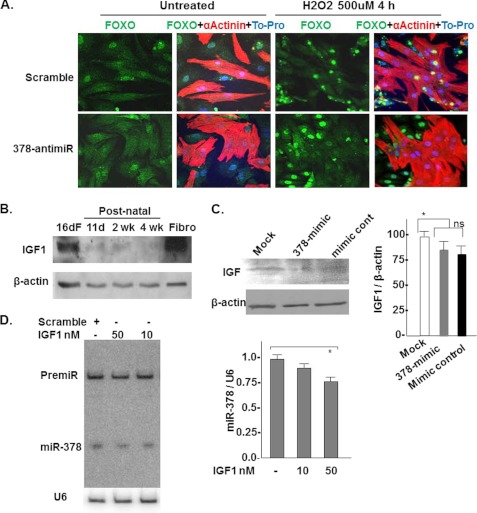
Postnatal cardiac remodeling is characterized by a marked decrease in the insulin-like growth factor 1 (IGF1) and IGF1 receptor (IGF1R) expression. The underlying mechanism remains unexplored. This study examined the role of microRNAs in postnatal cardiac remodeling. By expression profiling, we observed a 10-fold increase in miR-378 expression in 1-week-old neonatal mouse hearts compared with 16-day-old fetal hearts. There was also a 4-6-fold induction in expression of miR-378 in older (10 months) compared with younger (1 month) hearts. Interestingly, tissue distribution analysis identified miR-378 to be highly abundant in heart and skeletal muscles. In the heart, specific expression was observed in cardiac myocytes, which was inducible by a variety of stressors. Overexpression of miR-378 enhanced apoptosis of cardiomyocytes by direct targeting of IGF1R and reduced signaling in Akt cascade. The inhibition of miR-378 by its anti-miR protected cardiomyocytes against H(2)O(2) and hypoxia reoxygenation-induced cell death by promoting IGF1R expression and downstream Akt signaling cascade. Additionally, our data show that miR-378 expression is inhibited by IGF1 in cardiomyocytes. In tissues such as fibroblasts and fetal hearts, where IGF1 levels are high, we found either absent or significantly low miR-378 levels, suggesting an inverse relationship between these two factors. Our study identifies miR-378 as a new cardioabundant microRNA that targets IGF1R. We also demonstrate the existence of a negative feedback loop between miR-378, IGF1R, and IGF1 that is associated with postnatal cardiac remodeling and with the regulation of cardiomyocyte survival during stress.


Figures

FIGURE 1.
FIGURE 1.
Differential expression of microRNAs in postnatal heart. A, expression of randomly selected microRNAs in fetal (16-day gestation) and neonatal (7-day postnatal) mouse hearts by quantitative RT-PCR, asterisks mark p < 0.05 when compared with the fetal heart. B, validation of real-time PCR data by Northern analysis. C, schematic presentation and location of miR-378 in the first intron of the Pgc1β gene, stem-loop structure of pre-miR-378 and its processing into miR-378 and miR-378*. D, tissue distribution and development analysis of miR-378 and miR-378* by Northern blot analysis. E, expression of miR-378 in primary cultures of cardiomyocyte and fibroblasts obtained from the same culture (20 μg of RNA/lane). Each experiment was repeated a minimum of 3 times. U6 was used as the loading control.
FIGURE 2.
FIGURE 2.
miR378 is a stress inducible microRNA in rat neonatal cardiomyocytes. A, developmental and age-related expression of miR-378 by Northern blotting. B, expression of miR-378 following treatment of neonatal cardiomyocytes with various stressors for the indicated times and durations. Lower panel shows the expression of a non-related microRNA, miR-208, in the same membrane after stripping and hybridization with radiolabeled miR-208. C, quantification of miR-378 normalized to miR-208a, each bar is the mean ± S.D. of a minimum of 3 independent experiments. D, real-time PCR analysis of Pgc1β mRNA in the same samples as in B. Relative expression was calculated by the ΔΔCT method using β-actin as a reference gene. Asterisks mark the statistical significance (p < 0.05). NS, non-significant.
FIGURE 3.
FIGURE 3.
IGF1R as a predicted target of miR-378. A, sequence alignment of IGF1R 3′ UTR with the miR-378 seed sequence (highlighted in the box) showing the position of the predicted binding site and species conservation. B, Western blot analysis of IGF1R in the fetal heart and after birth (50 μg of protein/lane). C, inverse expression pattern of miR-378 and IGF1R protein levels in cardiac tissues at the indicated development time. Blots are representative of a minimum of 3.
FIGURE 4.
FIGURE 4.
miR-378 reduces endogenous IGF1R expression by direct targeting of 3′ UTR. A, Northern analysis showing increased expression of mature miR-378 48 h following transfection of the 378-mimic in cardiac myocytes, U6 was used as a loading control. B, IGF1R expression by Western blot in triplicate (50 μg of protein/lane) in the presence of mimic control or increasing amounts of 378-mimic, GAPDH used as a loading control. Right graph is derived from 3 additional experiments. C, real-time PCR analysis of IGF1R mRNA levels in mimic control and 378-mimic transfected cells (n = 2). D, functional assay of the IGF1R 3′ UTR in H9C2 cells using a dual luciferase reporter system following transfection of various DNA constructs in the presence of 378-mimic or mimic control. The sequence shown is the predicted target sequence of IGF1R 3′ UTR, three repeats of this sequence (WtIGF1R3X-luc) or mutated sequence (underlined nucleotide mutIGF1R3X-luc) were cloned downstream of the luciferase reporter, Renilla luciferase activity was used for normalizing data. Data are derived from triplicate transfectants of 3 independent experiments. *, p < 0.05.
FIGURE 5.
FIGURE 5.
Inhibition of IGF1 but not PE-induced signaling and augmentation of PQ-401 effect on pAkt by miR-378. A, Western analysis showing pAkt (Thr308) and total Akt in neonatal rat cardiomyocytes following transfection with mimic control or 378-mimic for 48 h, cells were then serum starved overnight and treated with IGF1 (10 nm) for 15 min. B, same cell lysates as in A analyzed for pERK and total ERK2. C, cells were prepared as in A, but treated with phenylephrine (50 μm) for 5 min and analyzed for pERK and total ERK. D, cells were prepared as in A, but treated with PQ-401 (10 μm) 90 min prior to stimulation with IGF1. All results are representative of at least three independent experiments. p < 0.05, Significant when compared with the non-treated group (*) or when compared with the corresponding treated mimic control group (#).
FIGURE 6.
FIGURE 6.
Effect of miR378 on cell survival. A, immunofluorescence images of cardiomyocytes 72 h after either mock transfection (control), mimic control or 378-mimic, and following a 4-h treatment with H2O2 (500 μm) showing ToPro-stained nuclei (blue), after TUNEL staining (green), and the two images merged together. Arrows mark the nuclei considered as TUNEL positive. B, quantification of TUNEL-positive nuclei as described for the different treatment groups. C, caspase 3/7 activity in cardiomyocytes transfected as in A and treated with H2O2 as indicated. #, significantly different from non-treated group; *, significant when compared with mock or mimic control transfected groups treated with H2O2 for the same period. D, Western analysis of IGF1R and signaling cascade in cardiomyocytes transfected as in A and after a 4-h H2O2 treatment. E, quantification of the signal intensity of D. Expression levels of IGF1R, pIGF1R, Bim, Trail, and FasL were normalized with β-actin, whereas pAkt and pFoxO3 were normalized with their non-phospho counterparts. *, p < 0.05. Each bar is a mean ± S.D. of a minimum of 3 independent experiments.
FIGURE 7.
FIGURE 7.
miR-378 knockdown enhances IGF1R expression and IGF1-induced AKT activation. A, Northern blot showing knockdown of miR-378 in cardiac myocytes 48 h after transfection with either scramble control or 378-anti-miR, U6 represents the loading control on the same membrane. B, Western analysis of IGF1R 72 h after transfection of cardiomyocytes with scramble or increasing amounts of 378-anti-miR. C, expression of pAkt and total Akt in cardiomyocytes transfected with scramble control or 378-anti-miR. After a 48-h transfection, cells were incubated in serum-free media overnight and then treated with IGF1 for 15 min. D, cells were prepared as in C and treated with the IGF1R inhibitor PQ-401 (10 μm) 90 min prior to IGF1 treatment. p < 0.05, *, compared with non-PQ treated scramble control; #, compared with the PQ-401-treated scramble control (n = 2).
FIGURE 8.
FIGURE 8.
miR-378 knockdown prevents the H2O2-induced apoptosis program and hypoxia/reoxygenation-induced cell death in an IGF1R-dependent manner. A, Western analysis (duplicates) of pIGF1R, IGF1R, and downstream signaling cascade in response to oxidative stress following transfection with either scramble control or 378-anti-miR. The same membrane was used again and again for probing with different antibodies after stripping. B, quantification of the signal intensity of A normalized essentially as described in the legend to Fig. 6E. C, time course response of TUNEL-positive nuclei in response to varying periods of hypoxia/reoxygenation in the presence of either scramble control or 378-anti-miR (50 nm). Each bar is a mean ± S.D. of a minimum of 3 independent experiments. D, quantification of TUNEL positive nuclei as described for the indicated treatment groups after 2 h of hypoxia followed by 2 h of reoxygenation injury to cardiac myocytes. The IGF1R inhibitor PQ-401(PQ) produced a dose-dependent inhibition of protective effects of 378-anti-miR. E, 378-anti-miR enhanced cardiomyocyte viability. Significant p < 0.05 (*) when compared with scramble control group and (#) when compared with the scramble PQ-treated group.
FIGURE 9.
FIGURE 9.
miR-378 inhibition interferes with FoxO3 translocation in the cardiomyocyte nuclei. IGF1 is a negative regulator of miR378. A, confocal imaging showing FoxO3 subcellular distribution in co-cultures of neonatal cardiomyocyte and non-muscle cells 72 h after transfection with scramble control or 378-anti-miR with and without H2O2 treatment for 4 h. FoxO3 can be visualized as green immunofluorescence, nuclei as blue, myocytes as red immunofluorescence α-actinin positive cells, and non-muscle cells as absence of red fluorescence. Note, with 378-anti-miR transfection and H2O2 treatment FoxO3 was excluded only from the nuclei (seen as purple-stained nuclei in the merged image) of red-stained muscle cells, whereas its presence was detected in the nuclei of the scramble control transfected muscle cells (seen as intense yellow staining in the merged image). B, IGF1 expression and time course by Western blot analysis in fetal and postnatal heart tissues and in cultured neonatal cardiac fibroblasts. C, miR-378 overexpression does not significantly affect IGF1 expression in cardiomyocytes. D, IGF1 acts as a negative regulator of miR-378. Primary cultures of cardiomyocytes were treated with increasing doses of IGF1 for 72 h. miR-378 expression was analyzed by Northern. A modest but significant reduction was noted with a higher dose of IGF1 (n = 2).

Similar articles

Cited by

References

    1. Lewis B. P., Burge C. B., Bartel D. P. (2005) Conserved seed pairing, often flanked by adenosines, indicates that thousands of human genes are microRNA targets. Cell 120, 15–20 - PubMed
    1. Berezikov E., Guryev V., van de Belt J., Wienholds E., Plasterk R. H., Cuppen E. (2005) Phylogenetic shadowing and computational identification of human microRNA genes. Cell 120, 21–24 - PubMed
    1. Small E. M., Olson E. N. (2011) Pervasive roles of microRNAs in cardiovascular biology. Nature 469, 336–342 - PMC - PubMed
    1. Chen J. F., Murchison E. P., Tang R., Callis T. E., Tatsuguchi M., Deng Z., Rojas M., Hammond S. M., Schneider M. D., Selzman C. H., Meissner G., Patterson C., Hannon G. J., Wang D. Z. (2008) Targeted deletion of Dicer in the heart leads to dilated cardiomyopathy and heart failure. Proc. Natl. Acad. Sci. U.S.A. 105, 2111–2116 - PMC - PubMed
    1. da Costa Martins P. A., Bourajjaj M., Gladka M., Kortland M., van Oort R. J., Pinto Y. M., Molkentin J. D., De Windt L. J. (2008) Conditional dicer gene deletion in the postnatal myocardium provokes spontaneous cardiac remodeling. Circulation 118, 1567–1576 - PubMed

Publication types

MeSH terms

LinkOut - more resources