Review
. 2012 Apr 4;31(7):1630-43.
doi: 10.1038/emboj.2012.42. Epub 2012 Feb 24.

Protease signalling: the cutting edge

Affiliations
Review

Protease signalling: the cutting edge

Boris Turk et al. EMBO J. .

Abstract

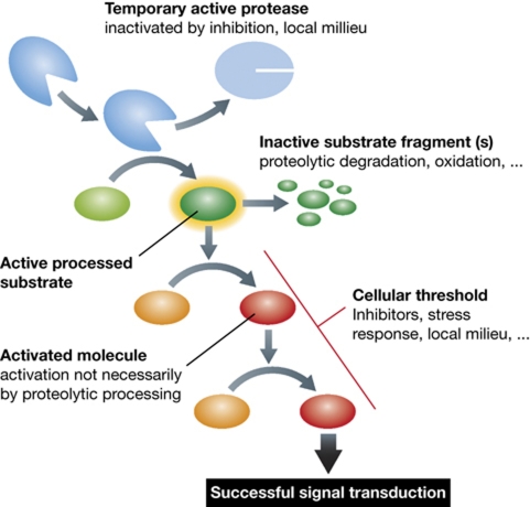
Protease research has undergone a major expansion in the last decade, largely due to the extremely rapid development of new technologies, such as quantitative proteomics and in-vivo imaging, as well as an extensive use of in-vivo models. These have led to identification of physiological substrates and resulted in a paradigm shift from the concept of proteases as protein-degrading enzymes to proteases as key signalling molecules. However, we are still at the beginning of an understanding of protease signalling pathways. We have only identified a minor subset of true physiological substrates for a limited number of proteases, and their physiological regulation is still not well understood. Similarly, links with other signalling systems are not well established. Herein, we will highlight current challenges in protease research.


Conflict of interest statement

The authors declare that they have no conflict of interest.

Figures

Figure 1
Figure 1
Binding of a substrate to a protease. (A) Schematic representation of substrate binding. The basic differences between binding of a small peptidic or peptidomimetic substrate and a larger peptide or protein substrate are shown. Small substrate binds tightly to the non-prime binding sites whereas the usually bulky fluorophore/chromophore (leaving group) binds to the S1′ site while the other prime sites remain empty (1). Larger peptidic substrates bind to both prime site and non-prime site binding sites, while the interaction with some individual binding sites can be looser (2). Exosites on protease surface serve as additional binding elements for large substrates to strengthen the interaction with the protease and allow recognition (3). They can be also used for discrimination between the different substrates. (B) Small substrate-mimicking inhibitors that helped tremendously in elucidating the substrate binding mechanism(s) often bind the same way as the small substrates (some more differences with metalloproteases), except that in covalent inhibitors the leaving group is replaced by a warhead (see A for a schematic representation). Crystal structure of caspase-1 in complex with the inhibitor YVAD-CHO (Wilson et al, 1994; 1ICE) revealed the insight into the mechanism of substrate binding to the active site of caspases as an example of such small molecule (substrate or inhibitor) binding. The surface of caspase 1 is shown in light grey. In the active site cleft, the non-primed and primed parts are shown as cyan and blue surface. The structure of the inhibitor (shown as a ball-and-stick model) has revealed the S1, S2, and S3 substrate binding sites. The non-hydrogen atoms C, N, and O are shown in blue, dark blue, and red, respectively. Figure was prepared with MAIN (Turk, 1992) and rendered with POV-Ray.
Figure 2
Figure 2
Protease activation. (A) Schematic representation of protease zymogen activation with proteases where active site is preexisting (a) or non-existing (b). (1) Propeptide removal in proteases with preexisting active site such as seen in cysteine cathepsins (see B as an example); (2) allosteric or conformational activation of proteases with distorted active site, such as chymotrypsin-like serine proteases (granzymes and trypsin), and some caspases; (3) conformational rearrangement and formation of the active site upon cofactor or platform-based activation such as seen in caspases and during Factor VII activation. (B) Crystal structure of a zymogen form of a protease offers a major support in understanding the mechanism of zymogen activation. Procathepsin B is shown as an example of a protease with preexisting active site. Propeptide region of cysteine cathepsins covers the active site cleft of the enzyme thereby blocking access to substrates. The mature part of procathepsin B (Podobnik et al, 1997; 3PBH) is shown as a white surface, whereas the active site residues C29 and H199 areas are in yellow and green, respectively. The propeptide is shown as a blue ribbon with the autocatalytic cleavage site (M56-F57) marked with an arrow. Figure was prepared with MAIN (Turk, 1992) and rendered with POV-Ray.
Figure 3
Figure 3
Inhibitors, major regulators of protease activities. (A) Schematic representations of different modes of inhibitor binding (adapted from Bode and Huber, 2000). (1) Substrate-like binding with direct blockade of the protease active site, such as seen with most inhibitors of trypsin-like serine proteases (see B for trypsin–antitrypsin interaction as an example); (2) steric blocking of the active site such as seen with a number of cathepsin inhibitors (see C for stefin A–cathepsin H interaction as an example); (3) exosite binding such as seen with a number of thrombin inhibitors and in a more extreme case with XIAP binding to caspases-3 and -7, which is a combination of sterical blocking of the active site and exosite binding; (4) quasi-substrate-like binding such as seen with TIMP binding to MMPs; (5) allosteric inhibition through distortion of the active site such as seen with propeptides of chymotrypsin-like proteases. (B) Inhibition of serine proteases by a serpin as an example of substrate-like binding with direct blockade of the protease active site (A1). The crystal structure of antitrypsin is shown in complex with trypsin (Huntington et al, 2000; 1EZX) is shown as an example. In the first step of inhibition, the RSL of the serpin is cleaved and the P1 residue M358 remains covalently bound to the reactive site S195 of trypsin. In the next step, 15 residues of the reactive loop (orange strand) carrying trypsin are inserted into the middle of the β-sheet of the serpin molecule (blue surface), resulting in a movement of the trypsin molecule (yellow surface) from the top to the bottom of the serpin. Trypsin catalytic site is thereby distorted and partially unfolded. The inserted part of the RSL of serpin and helix F were removed from the serpin surface in order to make the insertion visible behind the scaffold of the helix F. The ester link atoms between the side chain trypsin S195 and carbonyl group serpin M358 are shown in ball-and-stick representation. (C) Inhibition of cysteine cathepsins by endogenous inhibitors. The crystal structure of stefin A in complex with cathepsin H (Jenko et al, 2003; 1NB3) is shown as an example of steric blocking of the active site (A2). The folds of stefin A and cathepsin H are shown in ribbon presentation. Stefin A is on the top and is shown in yellow, while cathepsin H is shown in cyan. The mini-chain of cathepsin H is shown as a red stick model, while the three visible carbohydrate rings are shown in blue.
Figure 4
Figure 4
The apoptotic cascade. The two major apoptotic pathways, the extrinsic one (the death receptor pathway) and the intrinsic one (the mitochondrial pathway) are shown schematically. All the major proteolytic (black) and non-proteolytic steps (magenta) are marked with arrows, whereas the proteases (caspases and cathepsins) are shown in white characters in dark grey field. The lysosomal amplification loop is also marked, although the link between mitochondria and lysosomes has not been established yet. The direct destabilization of lysosomes (marked with a lightning) seems the only way that the lysosomes destabilization can trigger the apoptotic cascade, although it also ends up in self-amplification through the engagement of the mitochondrial pathway.
Figure 5
Figure 5
Signal transduction in protease signalling. A hypothetical signal transduction pathway is shown with the first step being a proteolytic processing of its substrate leading to the activation of the latter. This triggers a chain reaction of events, leading to transfer of the signal. However, if the cellular or reaction threshold (inhibitors, autophagy, …) is not overcome, the signal is not transmitted. If a protease is inactivated/degraded during the process, the signal can be transmitted if the overall threshold is overcome. Similar is true for subsequent processing/degradation or milieu-mediated inactivation of the protease substrate(s).

Similar articles

Cited by

References

    1. Acehan D, Jiang X, Morgan DG, Heuser JE, Wang X, Akey CW (2002) Three-dimensional structure of the apoptosome: implications for assembly, procaspase-9 binding, and activation. Mol Cell 9: 423–432 - PubMed
    1. Al-Khunaizi M, Luke CJ, Askew YS, Pak SC, Askew DJ, Cataltepe S, Miller D, Mills DR, Tsu C, Bromme D, Irving JA, Whisstock JC, Silverman GA (2002) The serpin SQN-5 is a dual mechanistic-class inhibitor of serine and cysteine proteinases. Biochemistry 41: 3189–3199 - PubMed
    1. Alvarez VE, Kosec G, Sant’Anna C, Turk V, Cazzulo JJ, Turk B (2008) Autophagy is involved in nutritional stress response and differentiation in Trypanosoma cruzi. J Biol Chem 283: 3454–3464 - PubMed
    1. Asagiri M, Hirai T, Kunigami T, Kamano S, Gober HJ, Okamoto K, Nishikawa K, Latz E, Golenbock DT, Aoki K, Ohya K, Imai Y, Morishita Y, Miyazono K, Kato S, Saftig P, Takayanagi H (2008) Cathepsin K-dependent toll-like receptor 9 signaling revealed in experimental arthritis. Science 319: 624–627 - PubMed
    1. Bar-Shavit R, Kahn A, Wilner GD, Fenton JW II (1983) Monocyte chemotaxis: stimulation by specific exosite region in thrombin. Science 220: 728–731 - PubMed

Publication types

Substances